
Sholto David uncovered yet another antibody cock-up! And again, the main problem is not just scientists using a completely unsuitable antibody, but the beautiful and convincing results they manage to produce nevertheless! And it is not just some Chinese labs, even one of most influential anti-aging researchers in USA is affected!
The antibody kerfuffle now is about β-galactosidase. Apparently the mammalian senescence-associated β-galactosidase and bacterial β-galactosidase are basically the same thing, because scientists have spoken.
I added a Coda about one of these US geniuses, to show you how exactly they manage to achieve perfect figures for publication in top journals without achieving any experimental data.
Here is Sholto’s past reporting about an antibody mix-up, it was about two very unrelated p16 proteins, which will also feature in this post again:

Mind over Antibody
“I considered whether I should email Abcam and ask them to change the name of the ab51243 antibody on their website to reduce confusion, but […] I will instead continue to watch the carnage, perhaps it has only just started!” – Sholto David

Caught Lac(Z)ing: Another Senescence Antibody Fumble
by Sholto David
Recently I wrote about a muddle in the ageing and senescence literature: Dozens of research teams have confused two proteins with similar names and ended up using an incorrect antibody for p16-INK4a. Shortly after that blog was published I was contacted by someone with a link to a 2016 Cell paper and a forgotten PubPeer thread which set off the second act of the senescence antibody farce.
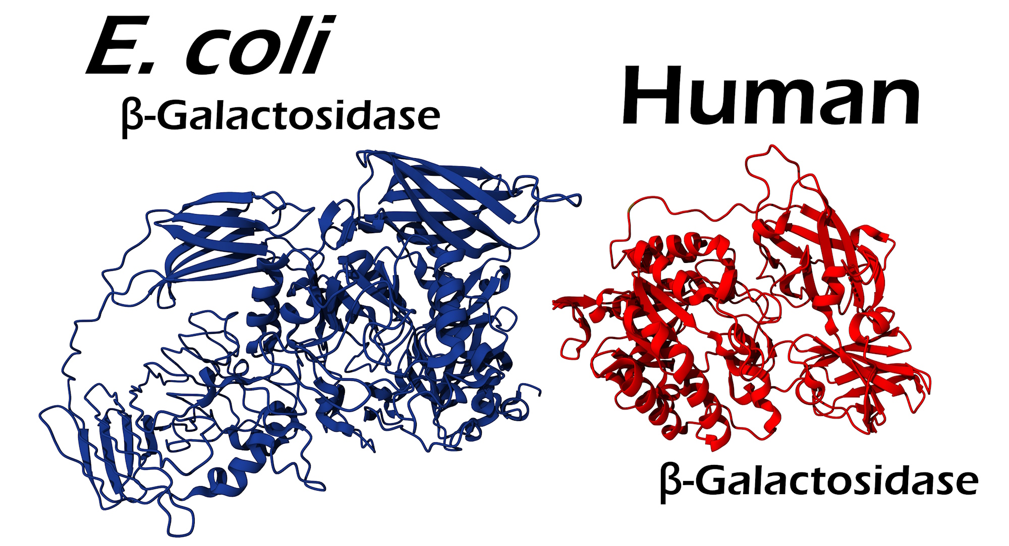
I’ll start with a little background… A useful observation about old (or “senescent”) cells is that they turn blue when exposed to the organic compound X-Gal. This blue colour is caused by the enzyme β-galactosidase which cleaves the otherwise colourless X-Gal leaving a nice blue precipitate. I’m not sure if there is a consensus for why old cells turn blue and young cells do not, but β-galactosidase is a lysosomal enzyme and there’s an idea that senescent cells acquire extra lysosomes – and that’s good enough for this blog.
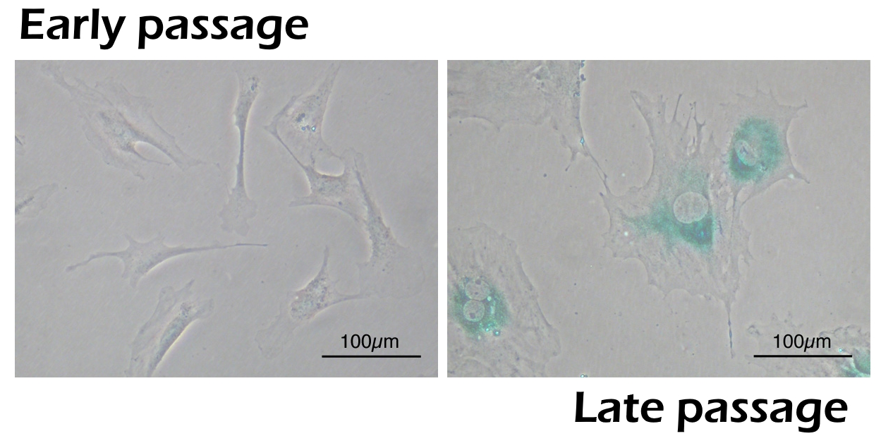
But what if you wanted to detect senescent cells without X-Gal? Well, some people have pursued an alternative strategy where they use an antibody to bind the β-galactosidase enzyme directly. With this method β-galactosidase can be visualised with immunostaining or western blotting. Without meaning to touch off a can of senescence worms it’s not quite clear whether this secondary technique truly marks senescent cells, but let’s just say it is a method with at least some scientific logic but perhaps not much validity (maybe that describes half the field of senescence, but I digress).
Now is a good time to note that the bacterium E. coli also has a famous β-galactosidase enzyme encoded by the LacZ gene, the discovery of which was rewarded with the Nobel Prize in 1965 (in mammals the gene is GLB1). The interaction of this E. coli enzyme with X-Gal has also found a practical use in various implementations of the LacZ reporter system. This genetics tool is used to monitor the activation of genes by expressing E. coli β-galactosidase under the control of specific promoters (usually in animal cells and tissue), allowing the visualisation of gene activation with the previously mentioned blue colour change.
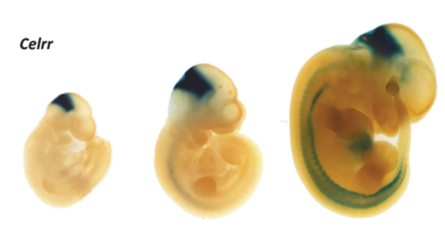
In cases where the colour change is not sufficiently bold or does not provide enough resolution, some labs also use an anti E. coli β-galactosidase antibody to probe for protein expression, again this can be done by immunostaining or western blotting.
So in summary, the endogenous mammalian β-galactosidase protein is used as a marker for senescent cells, while the E. coli β-galactosidase protein is a component of the versatile LacZ reporter system used for genetics experiments. While these enzymes have the same name, catalyse the same reaction, and share a useful substrate in X-Gal, they are otherwise related to separate lines of scientific inquiry.
So here is where the muddle happened: Some senescence researchers, when trying to stain or blot mammalian β-galactosidase as a marker of senescence, have been accidentally using antibodies specific to the E. coli β-galactosidase enzyme. This is a big blunder, using the wrong antibody completely invalidates the experimental results.
[Some people may ponder if perhaps there remains enough sequence homology between the mammalian and E. coli β-galactosidase enzymes for an antibody to bind both… the short answer is no, the long answer is at the end of this blog]
But for now, let’s look at some examples, we will start with a paper in Cell which I was initially sent, authored by the celebrity anti-aging researcher Juan Carlos Izpisua Belmonte, professor at Salk Institute, and founding director of Altos Labs institute in California, USA.

The Island of Dr Izpisua Belmonte
Human-monkey chimeras arrive to solve the problem of organ shortage. Thank Juan Carlos Izpisua Belmonte, who is ready to cure all possible diseases and even the old age. With chutzpah and Cell on his side.

Here is Figure 5E showing immunostaining of β-galactosidase in mouse liver tissue.
Alejandro Ocampo, Pradeep Reddy, Paloma Martinez-Redondo, Aida Platero-Luengo, Fumiyuki Hatanaka, Tomoaki Hishida, Mo Li, David Lam, Masakazu Kurita, Ergin Beyret, Toshikazu Araoka, Eric Vazquez-Ferrer, David Donoso, Jose Luis Roman, Jinna Xu, Concepcion Rodriguez Esteban, Gabriel Nuñez, Estrella Nuñez Delicado, Josep M. Campistol, Isabel Guillen, Pedro Guillen, Juan Carlos Izpisua Belmonte In Vivo Amelioration of Age-Associated Hallmarks by Partial Reprogramming Cell (2016) doi: 10.1016/j.cell.2016.11.052
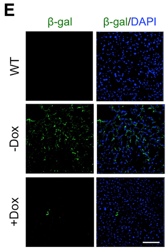
Figure 5 (E) Immunostaining of β-galactosidase in liver of LAKI 4F mice upon cyclic doxycycline administration.
The PubPeer discussion initially focused on whether immunostaining β-galactosidase as a senescence marker (instead of relying on the activity through X-Gal colour change) is a valid approach… The third comment mentions this critical piece of information:
“And just to add another detail. The antibody used in this paper, EMD Millipore #AB986, detects bacterial beta-galactosidase protein.”
Unregistered Submission, February 2017
Indeed, regardless of whether you think this approach of staining for β-galactosidase is valid, it will never work with an antibody raised against the E. coli β-galactosidase protein.
After I was sent this initial paper, I searched for the Millipore antibody and I couldn’t find any more examples of misuse. However, several other manufacturers sell antibodies against E. coli β-galactosidase, and plenty of those have been used for the wrong application, here is my list of antibody codes and the number of papers using these antibodies for the wrong application that I could find, so far 54 papers. There’s also a spreadsheet with further details linked here.
| Name | Supplier | Number of Wrong Papers |
| 14-6773-81 | ThermoFisher | 1 |
| A-11132 | ThermoFisher | 8 |
| ab116 | Abcam | 2 |
| ab616 | Abcam | 12 |
| ab4761 | Abcam | 3 |
| ab9361 | Abcam | 20 |
| AB986 | Millipore | 1 |
| sc-65670 | Santa Cruz | 7 |
Special prizes should be awarded to researchers who tried to probe for β-galactosidase and p16-INK4a (subject of my last blog) with wrong antibodies for both. Here’s a team from China, specifically from the Second Xiangya Hospital of Central South University:
Pan Tan, Yong-Hong Guo, Jun-Kun Zhan, Li-Min Long, Mei-Li Xu, Ling Ye, Xin-Yu Ma, Xing-Jun Cui, Hai-Qin Wang LncRNA-ANRIL inhibits cell senescence of vascular smooth muscle cells by regulating miR-181a/Sirt1 Biochemistry and cell biology (2019) doi: 10.1139/bcb-2018-0126

The authors obviously intended to blot senescence related proteins: “SA-β-Gal” is a slightly anachronistic abbreviation for “senescence associated β-galactosidase”. The antibodies listed for the western blot are ab9361 (specific to E. coli β-galactosidase) and ab51243 (specific to p16-ARC, not p16-INK4a). In fact Pan Tan and Hai-Qin Wang also made this same mistake again using the same wrong antibodies in a paper belonging to the Spandidos back-catalogue.
Pan Tan, Haiqin Wang, Junkun Zhan, Xinyu Ma, Xingjun Cui, Yanjiao Wang, Yi Wang, Jiayu Zhong, Youshuo Liu Rapamycin‑induced miR‑30a downregulation inhibits senescence of VSMCs by targeting Beclin1 International Journal of Molecular Medicine (2019) doi: 10.3892/ijmm.2019.4074

There are two more papers from different Chinese teams which include this double whammy mistake, one of them (from the First Affiliated Hospital of Hunan Normal University, ) doesn’t even bother to show the western blots, but does show quantitative analysis:
Hua Tang, Fang Yao, Ming Yin, Yangying Liao, Ke Li, Lan Li, Xiao Xiao, Junweichen Guo, Feng Hu, Hao Feng Anti-senescent effects of long non-coding RNA H19 on human dermal fibroblast cells through impairing microRNA-296-5p-dependent inhibition of IGF2 Cellular Signalling (2022) doi: 10.1016/j.cellsig.2022.110327

The second paper, from Nankai University in Tianjin, uses a different antibody for p16-ARC, ab220800, I hadn’t spotted this one when I wrote my last blog.
Yue Zheng, Yuchao Wang, Bingcai Qi, Wenqing Gao, Yanwu Liu, Tong Li Axin2 depletion in macrophages alleviated senescence and increased immune response after myocardial infarction Inflammation research (2023) doi: 10.1007/s00011-023-01843-8

Here’s an example from the lab of Dan Liebermann at Temple University in USA. The offending image was supposedly stained with ab616 – this is an antibody for the E. coli β-galactosidase enzyme but the authors intended to stain for human β-galactosidase. The image unexpectedly overlaps with the Phospho-JNK stained image… so perhaps the E. coli specific antibody didn’t perform as expected?
Jennifer S. Tront, Yajue Huang, Albert A Fornace Jr, Barbara Hoffman, Dan A Liebermann Gadd45a Functions as a Promoter or Suppressor of Breast Cancer Dependent on the Oncogenic Stress Cancer Research (2010) doi: 10.1158/0008-5472.can-10-2177
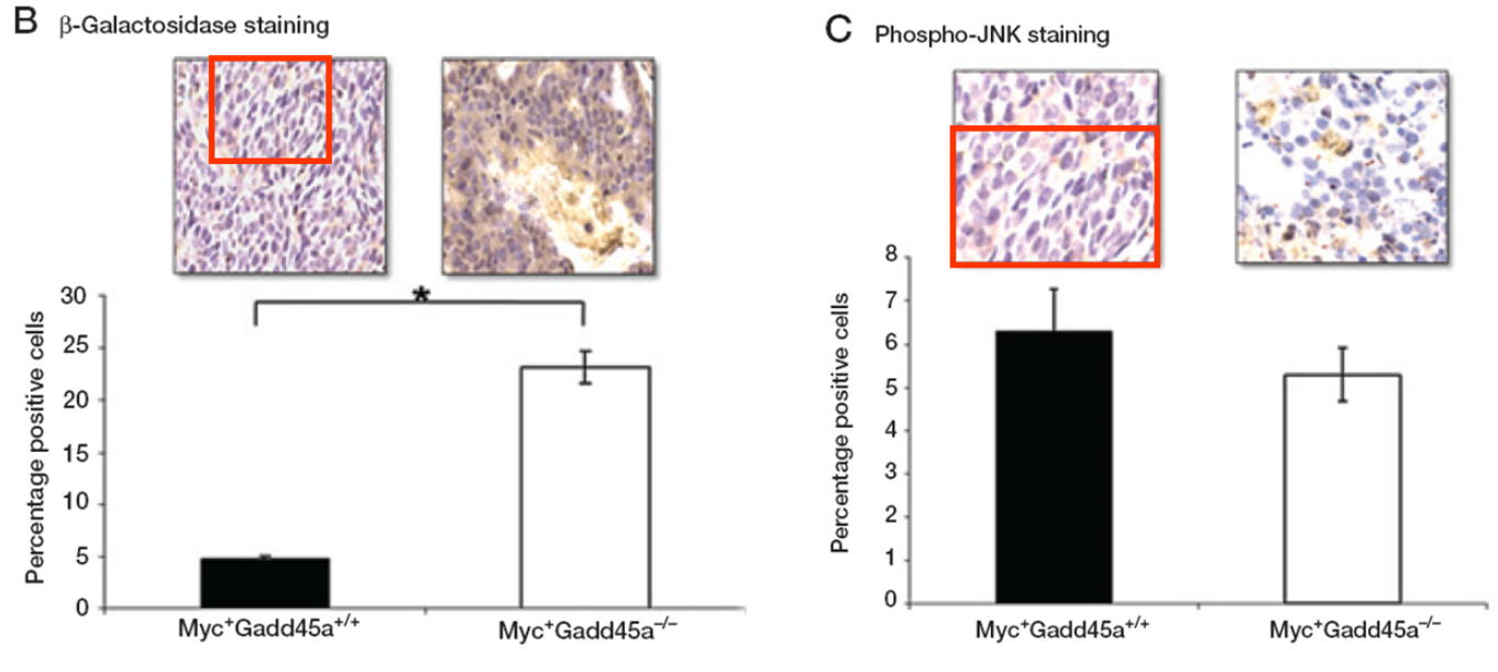
An image in Figure 3B and Figure 3C seem to overlap, with a different magnification and aspect ratio, but are described differently. Could the authors please check?
Unsurprisingly, this paper has other image irregularities, and yes, Liebermann is well aware of it since the last six years, and does nothing.

He has many more papers on PubPeer flagged for image forgeries, as you will see in the Coda at the end.
Some people have been quite frank about their findings, here a Chinese team puzzle about conflicting results from their efforts to stain β-galactosidase (bear in mind the paragraph doesn’t quite make sense, but this was published in a Nature family journal):
“Additionally, we compared cytoplasmic β-galactosidase (β-gal), a widely used marker of cell senescence, between control and 3xDR microglia. However, we did not observe β-gal elevation in 3xDR microglia (Extended Data Fig. 7e). The conflicting results, although the ‘senescence index’ does not technically distinguish senescence and aging, thus complicate the landscape of the microglial senescence state.”
Li, Xiaoyu, et al 2023 doi: 10.1038/s43587-023-00479-x
Here’s the figure in question, perhaps the conflicting results were caused by the antibody used (A-11132) being specific to E. coli β-galactosidase? The purple staining is likely entirely non-specific. I am no expert in immunostaining but it doesn’t seem to be associated with any cellular structures.
Xiaoyu Li, Yuxin Li, Yuxiao Jin, Yuheng Zhang, Jingchuan Wu, Zhen Xu, Yubin Huang, Lin Cai, Shuai Gao, Taohui Liu, Fanzhuo Zeng, Yafei Wang, Wenxu Wang, Ti-Fei Yuan, Hengli Tian, Yousheng Shu, Feifan Guo, Wei Lu, Ying Mao, Xifan Mei, Yanxia Rao, Bo Peng Transcriptional and epigenetic decoding of the microglial aging process Nature Aging (2023) doi: 10.1038/s43587-023-00479-x

Here’s a fun paper about feeding rats mulberry extract in an effort to reduces oxidative stress in aging vasculature. The authors included SA-β-Gal staining of the rat’s aortas as Figure 1(a) so they must have thought it was important, but they also used an antibody specific for the E. coli β-galactosidase (ab9361). Curiously, I can’t see much correlation between the staining intensity in Figure 1(a) compared the quantitative analysis in Figure 1(b). An anonymous PubPeer user found that the figure includes some fakery too. Maybe that’s why the figure doesn’t make much sense?
Geum‐Hwa Lee, The‐Hiep Hoang, Eun‐Soo Jung, Su‐Jin Jung, Seong‐Kyu Han, Myoung‐Ja Chung, Soo‐Wan Chae, Han‐Jung Chae Anthocyanins attenuate endothelial dysfunction through regulation of uncoupling of nitric oxide synthase in aged rats Aging Cell (2020) doi: 10.1111/acel.13279


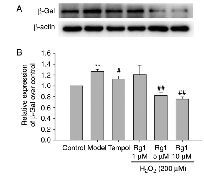
And if we’re going to take mulberries to prevent heart disease, we might as well throw in some ginseng to prevent Alzheimer’s, this time with Abcam’s ab9361 used in a western blot.
Tan‑Zhen Xu, Xiao‑Yan Shen, Ling‑Ling Sun, Ya‑Li Chen, Bi‑Qiong Zhang, Da‑Ke Huang, Wei‑Zu Li Ginsenoside Rg1 protects against H2O2‑induced neuronal damage due to inhibition of the NLRP1 inflammasome signalling pathway in hippocampal neurons in vitro International Journal of Molecular Medicine (2018) doi: 10.3892/ijmm.2018.4005
Not only academics fiddling around with plant extracts, but also serious industry teams with real drugs have muddled their β-galactosidase species. Here’s a well-cited paper in Oncotarget by Eli Lilly researchers based in Spain, this team has used Abcam’s ab9361 for a forbidden purpose…. No image here as the authors chose not to show the stained cells… I wonder why!?
Raquel Torres-Guzmán, Bruna Calsina, Ana Hermoso, Carmen Baquero, Beatriz Alvarez, Joaquín Amat, Ann M. McNulty, Xueqian Gong, Karsten Boehnke, Jian Du, Alfonso De Dios, Richard P. Beckmann, Sean Buchanan, María José Lallena Preclinical characterization of abemaciclib in hormone receptor positive breast cancer Oncotarget (2017) doi: 10.18632/oncotarget.17778

Where the authors did show image data, it turned out to be problematic:

David Goltzman is co-director of the Centre for Advanced Bone and Periodontal research at McGill University in Canada, and victim of harassment by cowardly PubPeer users:
David Goltzman’s cowardice
“It is cowardly to be anonymous. […] This is harassment, for whatever benefit you will gain from this. Please cease and desist” – David Goltzman
Goltzman has made many ill-fated collaborations with a certain Dengshun Miao from at Nanjing Medical University in China, an owner of a scary PubPeer record of almost 70 papers. Two of their papers used Abcam’s E. coli β-galactosidase antibody ab616 to stain for mouse β-galactosidase. Interestingly in the 2020 paper, the methods imply that western blotting was performed with this same antibody… although no western blot data was presented. A clue?
Haijian Sun, Wanxin Qiao, Min Cui, Cuicui Yang, Rong Wang, David Goltzman, Jianliang Jin, Dengshun Miao The Polycomb Protein Bmi1 Plays a Crucial Role in the Prevention of 1,25(OH)2D Deficiency‐Induced Bone Loss Journal of Bone and Mineral Research (2020) doi: 10.1002/jbmr.3921

Here the second paper, it also has a recycled western blot for p16 results:
Renlei Yang, Jiao Zhang, Jie Li, Ran Qin, Jie Chen, Rong Wang, David Goltzman, Dengshun Miaoa Inhibition of Nrf2 degradation alleviates age-related osteoporosis induced by 1,25-Dihydroxyvitamin D deficiency Free Radical Biology and Medicine (2022) doi: 10.1016/j.freeradbiomed.2021.12.010


Blogs about “irregularities” are hard to complete without at least one example from the field of long non-coding RNA; so here’s one with a classic formulaic title which presents western blots for β‐galactosidase throughout the paper (Figure 1, 3, 5, and 6) using Abcam’s ab9361. Of course, there is also an overlapping image.
Jiamei Yao, Zanhua Shi, Xinhua Ma, Daomiao Xu, Guangfeng Ming lncRNA GAS5/miR‐223/NAMPT axis modulates the cell proliferation and senescence of endothelial progenitor cells through PI3K/AKT signaling Journal of Cellular Biochemistry (2019) doi: 10.1002/jcb.28713


And I would never want to waste an opportunity to highlight the work of the “excellent team” at Scientific Reports. If there is a scientific stuff-up, they would never want to be left out.

Scientific Reports 2025: A Year in Review
“In this blog I write about papers published by Scientific Reports in 2025, so we could consider it to be a sort of “wrap-up” of highlights and special achievements in the world’s biggest scientific journal™ in 2025.” – Sholto David
Here, in the context of discussing senescence markers, a Japanese team comment that the expression level of β-galactosidase in their Caspase-3 knockout mice is higher, but the difference is not significant. Of course it might have been a more interesting result if they had used an antibody that would actually bind the right protein; once again this team used Abcam’s ab9361.
Takashi Ohnishi, Katsuhisa Yamada, Koji Iwasaki, Takeru Tsujimoto, Hideaki Higashi, Taichi Kimura, Norimasa Iwasaki, Hideki Sudo Caspase-3 knockout inhibits intervertebral disc degeneration related to injury but accelerates degeneration related to aging Scientific Reports (2019) doi: 10.1038/s41598-019-55709-3

Supplementary Figure S3 Higer β-galactosidase (β-gal) expression in nucleus pulposus (NP) cell colonies of caspase-3 knockout (KO) mouse compared with wild-type (WT) mouse
Here’s a recent paper in the journal Pain from one of the illustrious “Top Italian Scientists“, Nicoletta Galeotti, professor at University of Florence, this time using Santa-Cruz’s sc-65670, also clearly described as an E. coli specific antibody.
Vittoria Borgonetti, Nicoletta Galeotti Microglia senescence is related to neuropathic pain–associated comorbidities in the spared nerve injury model Pain (2023) doi: 10.1097/j.pain.0000000000002807

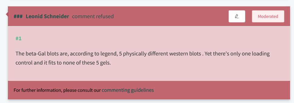
I want to finish by discussing what I expect will be a common response to this comment. Some people will insist that somehow, someway, these E. coli β-galactosidase specific antibodies can in fact bind to the endogenous mammalian β-galactosidase. In fact I have already received such a response from the authors of this eLife paper who used Abcam’s ab9361, one of them being the known cheater from Spain, Pura Muñoz-Cánoves:
Omid Mashinchian, Xiaotong Hong, Joris Michaud, Eugenia Migliavacca, Gregory Lefebvre, Christophe Boss, Filippo De Franceschi, Emmeran Le Moal, Jasmin Collerette-Tremblay, Joan Isern, Sylviane Metairon, Frederic Raymond, Patrick Descombes, Nicolas Bouche, Pura Muñoz-Cánoves, Jerome N Feige, C Florian Bentzinger In vivo transcriptomic profiling using cell encapsulation identifies effector pathways of systemic aging eLife (2022) doi: 10.7554/elife.57393
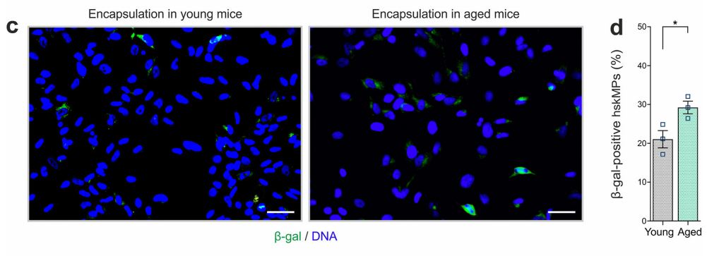
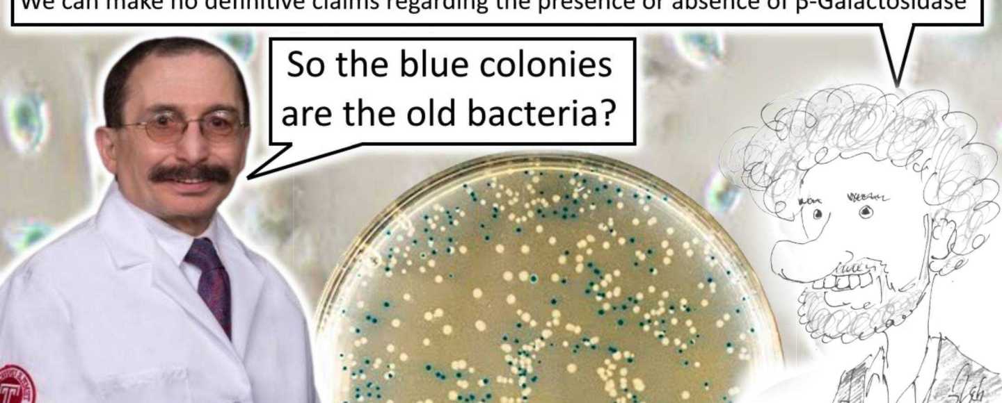
Florian Bentzinger, professor at University of Sherbrooke in Canada, also made this rather bemusing comment to me by email in June 2026:
“Importantly, our analyses were designed to compare relative signal between biological conditions under identical experimental settings, rather than to make definitive claims regarding the absolute presence or abundance of β-galactosidase itself.“
Florian Bentzinger
So when viewing an experiment using an anti β-galactosidase antibody and labelled as showing β-gal staining we should apparently not jump to any hasty conclusions and presume the authors actually claim the green staining is β-galactosidase. Always learning!

Wagner Ring Festival in Vienna
Erwin Wagner is not related to the famous German composer but we shall enjoy the Ring Festival with him and his friends anyway.
I will explore some lines of reasoning that should thoroughly refute this idea the antibodies really do cross react between species, but it will require some boring bioinformatics and even worse, looking at papers carefully.
Firstly, the sequence similarity between human and E. coli β-galactosidase is so low that it is not much better than random. Using Clustal Omega to align human β-galactosidase with E. coli β-galactosidase, the sequence identity is 20.1%. Replacing the human β-galactosidase sequence with a randomly generated sequence of the same length results in an alignment with a sequence identity of around 19% (using average amino acid composition from UniProtKB). For context, a common rule of thumb is that around 50% identity is required for a chance of antibody cross reactivity (with many exceptions of course, none of which apply here).
Another difficulty with this cross-species-binding argument, is that E. coli β-galactosidase has closer homologues in the human proteome than human β-galactosidase. If an antibody raised against the E. coli protein really does bind something in the human proteome, it’s far more likely to be human β-glucuronidase (25.0% identity) than human β-galactosidase (20.1% identity)… I’ve created a phylogenetic tree to illustrate this point…
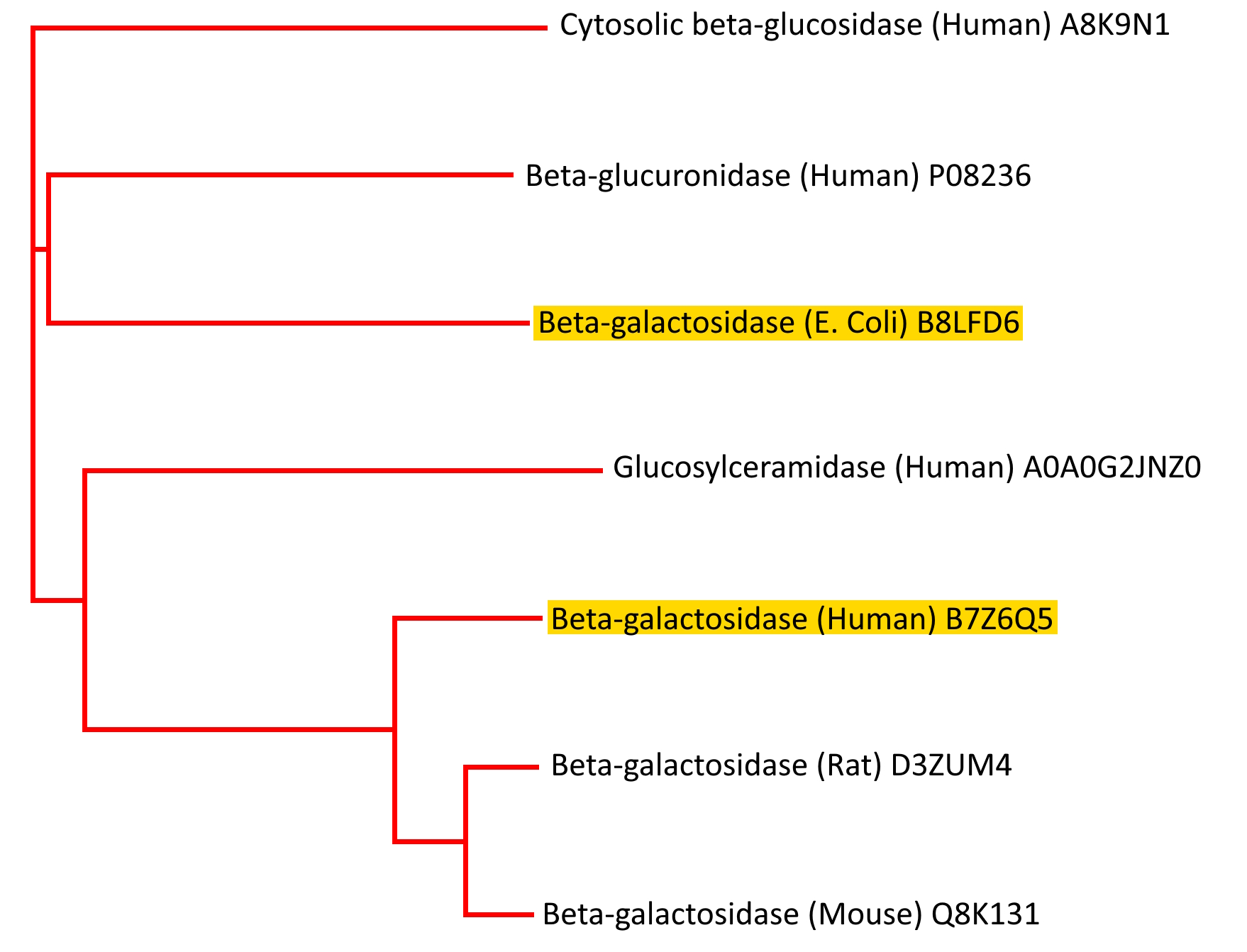
The tree shows that it is a false assumption to believe that the shared substrate specificity also indicates a common evolutionary thread (although all these enzymes do share a structural motif in the TIM barrel, it is a very widespread one, it’s unclear to me whether the TIM barrel has evolved more than once but I think we are exhausting patience by now).
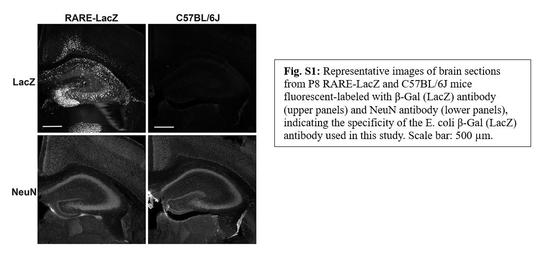
Apart from theory, there is also substantial experimental evidence that would refute this idea of cross-reactivity. Given the frequent use of the E. coli β-galactosidase enzyme in reporter gene systems, numerous papers report negative controls for LacZ expression staining, here’s a recent paper showing lack of background staining in mouse brain with ab9361.
Mariko Saito, Jungann Park, Anusha Nalluri, Brandon Marino, Colin R.O. Williams, Donald A. Wilson, Bhaskar C. Das, John F. Smiley Effects of ethanol exposure in neonatal mice on retinoic acid signaling in forebrain neurons and astrocytes IBRO Neuroscience Reports (2026) doi: 10.1016/j.ibneur.2026.05.006
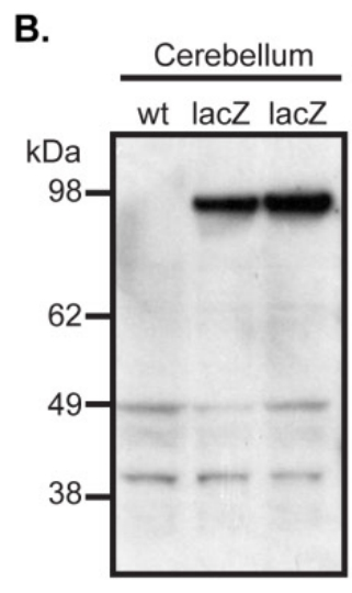
Here’s an example that shows the specificity of ThermoFisher’s A-11132 by western blot, note the lack of signal for b-galactosidase in the wild type column indicating the the antibody does not bind to the endogenous proteins but does bind to LacZ encoded β-galactosidase.
James L. J. Coleman, Margaret A. Mouat, Jianxin Wu, Nikola Jancovski, Jaspreet K. Bassi, Andrea Y. Chan, David T. Humphreys, Nadine Mrad, Ze-Yan Yu, Tony Ngo, Siiri Iismaa, Cristobal G. Dos Remedios, Michael P. Feneley, Andrew M. Allen, Robert M. Graham, Nicola J. Smith Orphan receptor GPR37L1 contributes to the sexual dimorphism of central cardiovascular control Biology of Sex Differences (2018) doi: 10.1186/s13293-018-0173-y
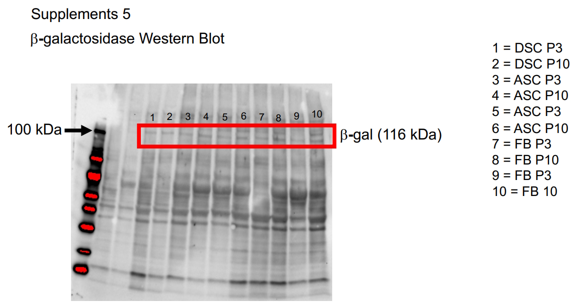
Finally, as noted above, several of the papers using these wrong antibodies seem to report results that the authors find hard to interpret. Here’s another interesting example where the authors try to blot for mammalian β-galactosidase with Abcam’s E. coli specific ab616. A full western blot is shown at the end of the paper although the purpose of the blot is unclear. Note that the molecular weight for E. coli β-galactosidase is highlighted at 116 kDa, although the authors intended to blot for human β-galactosidase (76 kDa). I think this western blot is probably an accurate representation of what you will actually see if you blot for the mammalian protein with the E. coli antibody, it looks like a non-specific mess and that’s probably why the authors didn’t even try to explain it, and published in Scientific Reports.
Vera Grotheer, Nadine Skrynecki, Lisa Oezel, Jan Grassmann, Joachim Windolf Osteogenic differentiation of human mesenchymal stromal cells and fibroblasts differs depending on tissue origin and replicative senescence Scientific Reports (2021) doi: 10.1038/s41598-021-91501-y
Note from LS: the last author of this paper used to be Christoph Suschek, a close friend of mine during my time as PhD student in Düsseldorf. Christoph told me now that he “withdrew from the list of authors because I was not even remotely satisfied with the results, the interpretation, and certainly not with the presentation“, and pointed me to two corrections of this paper (July 2021 and August 2021), which stated:
“Christopher V. Suschek was incorrectly listed as an author of the original Article, and has subsequently been removed”.
So to summarise my reply to the “it binds both proteins” response: The sequence identity is very low, if there really was any cross-species reactivity from an E. coli specific β-galactosidase antibody, it would probably be to human β-glucuronidase or another glucosidase, but not human β-galactosidase. The evidence in the literature seems to indicate this cross-reactivity does not occur (at best, the evidence is mixed). So if the authors of these papers wish to make that argument, that the antibody they have used binds proteins cross-kingdom, they would need some clearly presented experimental evidence with appropriate controls… Not found in any of these papers!
Besides – why in the world would anyone choose an E. coli specific antibody when there are plenty of mammalian specific antibodies for GLB1 encoded β-galactosidase on the market!? It makes no sense as a decision, and none of these papers try to explain it.
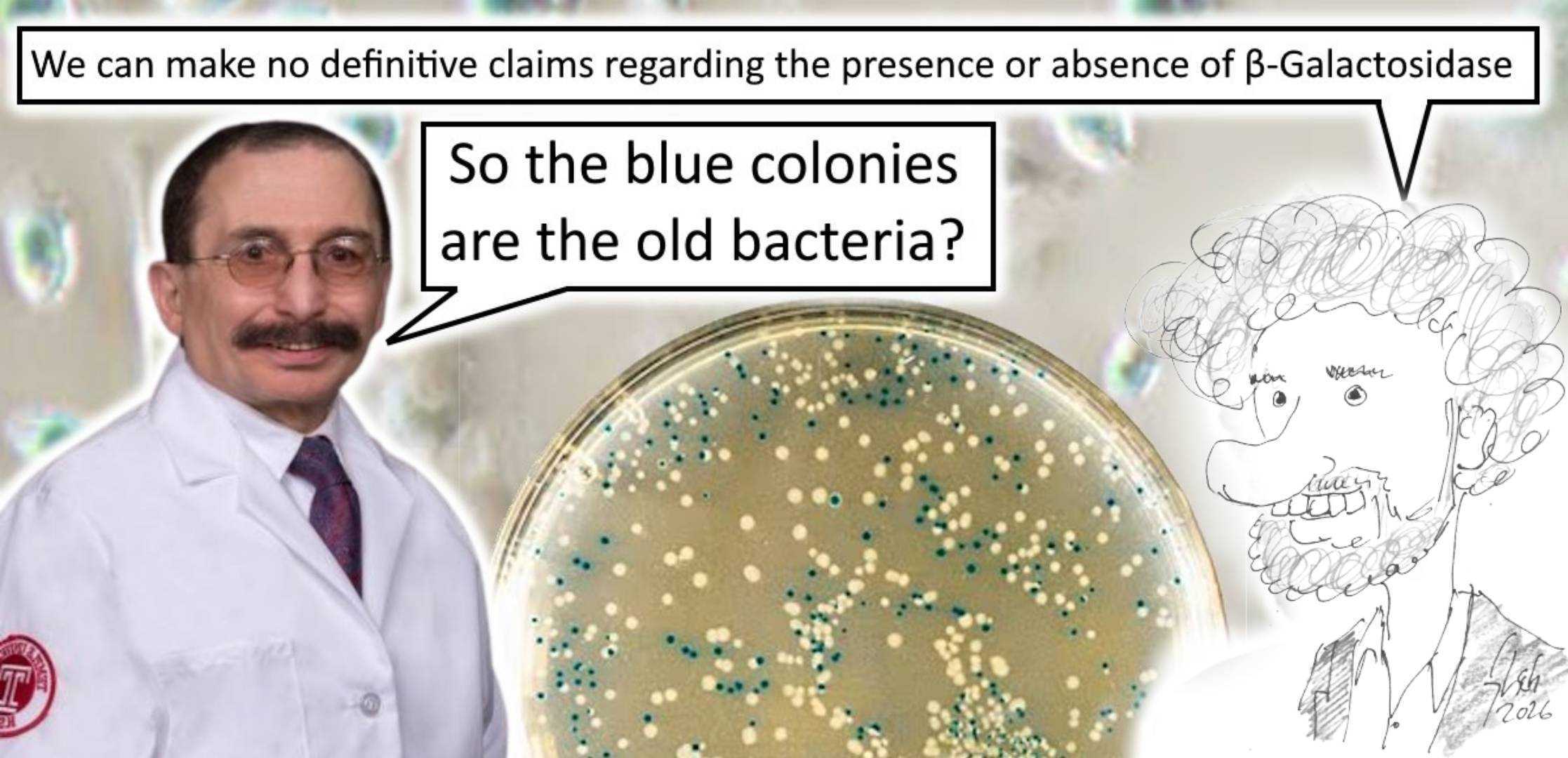
“I recently became aware that the article […] incorporates my photograph into its header graphic without my knowledge or consent. […] I do not consent to the use of my likeness in connection with this article. Accordingly, I respectfully request that my photograph be removed from the article and any associated promotional materials and replaced with an alternative image that does not depict me.”
We now complied and replaced the image of Prof Bentzinger with a cartoon of an unrelated generic scientist.
I’m not going to repeat all of my thoughts from my last discussion of antibody mix ups. It is unfortunate that such shenanigans occur unchecked when simple controls and peer-review should catch them quite quickly. Once again I am left wondering what to make of these research teams that cheerfully go on generating impossible results, either they don’t write the methods truthfully or they don’t write the results truthfully and neither one is better than the other.
Some people might be surprised that such a mistake is possible at all, but remember that there is an extensive and growing body of literature discussing the phenomena of so called “bacterial mitochondria” (also read September 2025 Shorts) – the depths of scientific stupidity are always expanding…
Zhaonan Hao, Mingbo Wang, Lin Cheng, Minmin Si, Zezhou Feng, Zhiyuan Feng Synergistic antibacterial mechanism of silver-copper bimetallic nanoparticles Frontiers in Bioengineering and Biotechnology (2024) doi: 10.3389/fbioe.2023.1337543

Any more antibody mix-ups? Let us know in the comments or by email! I already know of one more big one.


CODA by LS: The eternal art of Dan Liebermann
As promised, here is the Coda about Dan Liebermann, professor at Lewis Katz School of Medicine at Temple University in USA, who, as also promised, has many papers on PubPeer, currently 19 to be precise.
He also sports an enormous mustache – he probably was trying to look like the painter Max Liebermann, but overdid it:


What is also enormous, is the scale of image forgery in Liebermann’s papers. This fake paper is almost three decades old:
K Krishnaraju , B Hoffman, D A Liebermann The zinc finger transcription factor Egr-1 activates macrophage differentiation in M1 myeloblastic leukemia cells Blood (1998) doi: 10.1182/blood.V92.6.1957


Again, Liebermann is out for Blood:
A Amanullah , B Hoffman , D A Liebermann Deregulated E2F-1 blocks terminal differentiation and loss of leukemogenicity of M1 myeloblastic leukemia cells without abrogating induction of p15(INK4B) and p16(INK4A) Blood (2000) doi: 10.1182/blood.V96.2.475


And look at this artwork, like the others also rotting on PubPeer since 2017:
Marianna Shafarenko , Dan A. Liebermann , Barbara Hoffman Egr-1 abrogates the block imparted by c-Myc on terminal M1 myeloid differentiation Blood (2005) doi: 10.1182/blood-2004-08-3056


Impressed? Another one with Liebermann’s close associate at Temple University, Barbara Hoffman, in the same society-run journal:
Marianna Shafarenko, Arshad Amanullah, Bernard Gregory, Dan A. Liebermann, Barbara Hoffman Fos modulates myeloid cell survival and differentiation and partially abrogates the c-Myc block in terminal myeloid differentiation Blood (2004) doi: 10.1182/blood-2002-09-2704


Another ancient paper in another journal:
Wei Zhang, Insoo Bae, Kandasamy Krishnaraju, Naiyer Azam, Wen Fan, Kendall Smith, Barbara Hoffman, Dan A Liebermann CR6: A third member in the MyD118 and Gadd45 gene family which functions in negative growth control Oncogene (1999) doi: 10.1038/sj.onc.1202885

More bad gels by Liebermann and Hoffman:

Fig 4

Fig 1B

Fig 1

Fig 3A and 6D
Luckily, the Temple University not only doesn’t care about fraud, it shamelessly rewards it.

Steven Houser and the Temple of Fraud
“We all hype our work. We want to tell people our work is important. These patients, many of them coming to enroll in these trials, they have no other hope.” -Steven Houser, Hero of Research Ethics, Temple University
Also flow cytometry got fudged by the Liebermann-Hoffman duo:
Mamta Gupta, Shiv K Gupta, Arthur G Balliet, Mary Christine Hollander, Albert J Fornace, Barbara Hoffman, Dan A Liebermann Hematopoietic cells from Gadd45a- and Gadd45b-deficient mice are sensitized to genotoxic-stress-induced apoptosis Oncogene (2005) doi: 10.1038/sj.onc.1208847


This is just lazy:
D L Vesely , B Hoffman , D A Liebermann Phosphatidylinositol 3-kinase/Akt signaling mediates interleukin-6 protection against p53-induced apoptosis in M1 myeloid leukemic cells Oncogene (2007) doi: 10.1038/sj.onc.1210109

No wonder Liebermann got that false antibody to perform beautifully! All this lets me wonder if his enormous mustache is fake also?


Donate!
If you are interested to support my work, you can leave here a small tip of $5. Or several of small tips, just increase the amount as you like (2x=€10; 5x=€25). Your generous patronage of my journalism will be most appreciated!
€5.00


“CODA by LS: The eternal art of Dan Liebermann
As promised, here is the Coda about Dan Liebermann, professor at Lewis Katz School of Medicine at Temple University in USA, who, as also promised, has many papers on PubPeer, currently 19 to be precise.”
It might be a Philadelphia thing. Temple University is in Philadelphia, as is Thomas Jefferson University, where Calabretta, Bruno is.
Bruno Calabretta, 27 entries at Pubpeer. PubPeer – Search publications and join the conversation.
I can remember when 1999 was in the future. “What is also enormous, is the scale of image forgery in [Bruno Calabretta]’s papers. This fake paper is almost three decades old”: PubPeer – Activation of mitochondrial Raf-1 is involved in the antiapo…
I mean, it’s O.K. really, the image forgery in only in Cancer Research, sets the AACR (American Association for Cancer Research) standard.
Bruno Calabretta “out for Blood”, just like Dan Liebermann PubPeer – Leukemogenesis induced by wild-type and STI571-resistant BCR…
LikeLiked by 1 person
First, I wholeheartedly agree with the assessment that the antibodies are ill-equipped to analyze b-galactosidase. However, I want to bring up one counterpoint. Obviously most companies do not disclose the specific antigen they use for generating antibodies, but what if the sequence they used here was conserved between proteins? Wouldn’t that be more important than looking at the whole sequence homology? Regardless, you’d hope researchers would validate antibodies… how do you follow a whole research directions without even having the tools to evaluate??
LikeLike
What you are suggesting is impossible because there are no more than four consecutive amino acid residues that can be aligned between these two proteins, the longest common sequence is “DYLR”. If you could create an antibody against that four residue peptide it would also cross react to many other proteins in the human proteome and be useless. Although the exact sequence is not shared most do say something along the lines of “whole E. coli enzyme”, “40 amino acid sequence”. Here’s a link to an alignment between human and E. coli protein, it should last a few days: https://tcoffee.crg.eu/apps/tcoffee/result?rid=7de7d53a
LikeLike
Important to repeat that although you might think you can see some commonality in the alignment it is elusive – an alignment to randomly generated sequence of appropriate composition will look similar.
LikeLike
Ah, yes I was lazy and did not align sequences. Thanks!
LikeLike
As someone who worked in this field, I have a difficult time even imagining what use antibodies are in the first place. The marker was meant to be b-galactosidase *activity* in *acidic pH*, none of which are detected by antibodies in the first place.
And everybody knows this is bullshit to begin with. All cells have some b-gal activity and can be tortured into positivity by one of many means, including exposure time. Campisi, one of the popes who built this church of nonsense pseudoscience, did herself warn that the marker was meaningless in isolation.
LikeLiked by 2 people
Yes, several senescence researchers said the same: the antibody approach is dubious at best. Presumably people do this when torturing efforts are exhausted.
LikeLiked by 1 person
So how does this work, peer review-wise?
You reject papers where the antibody method was used, unless the authors are white?
LikeLike
Oh, there is more “bottom” of this particular barrel to scrape when you’re desperate. PCR for the b-gal gene, anyone?
LikeLiked by 2 people
In the absence of experimental evidence demonstrating that this antibody does not recognize its intended target, you are making very strong claims. More importantly, your discussion suggests a limited understanding of the biological factors that determine antibody recognition.
Primary amino acid sequence similarity is only one aspect that influences whether a polyclonal antibody will recognize a given protein. Structural conservation is often equally, if not more, important. For example, when conservative amino acid substitutions are taken into account, the sequence similarity between bacterial LacZ and murine or human GLB1 increases to approximately 35-40%. If one focuses on catalytically important regions, the similarity is even higher. Consequently, sequence identity alone is not sufficient grounds for dismissing the possibility of recognition or cross-reactivity.
It is also important to consider all existing literature, which you conveniently omit from your discussion. Multiple studies have validated the use of this antibody alongside established senescence markers. A quick google search shows for instance:
https://pmc.ncbi.nlm.nih.gov/articles/PMC7425512/
https://pubmed.ncbi.nlm.nih.gov/40311867/
Furthermore, the same antibody has been included in published protocols for endogenous GLB1 immunostaining in human tissues (e.g., https://pubmed.ncbi.nlm.nih.gov/27812879/). While no single publication constitutes definitive proof of specificity, the cumulative body of evidence should at least be considered before dismissing the reagent outright. Ignoring this literature while presenting a categorical conclusion gives an incomplete and potentially misleading picture of the available evidence.
All that being said, the way you approach this discussion falls far short of what one would expect from a serious scientific debate. Raising concerns about scientific misconduct when supported by clear evidence, such as image manipulation or data falsification, is entirely appropriate. However, making sweeping claims about the performance of an antibody without providing corresponding experimental evidence is a very different matter.
Your comics read less like a substantive scientific critique and more like a childish exercise in sensationalism aimed at attracting attention. Scientific conclusions are evaluated on the basis of evidence, experimental validation, and careful consideration of the relevant literature, not on confidence, rhetoric, or the selective presentation of information.
LikeLike
Hi Florian Bentzinger. Your IP address is in Sherbrooke, Canada.
Get a life.
LikeLiked by 2 people
It really is Florian, champion of open scientific debate hiding behind a fake name, and very likely augmented by ChatGPT, perhaps this is the most important line that you wrote:
“Your comics read less like a substantive scientific critique and more like a childish exercise in sensationalism aimed at attracting attention.”
LikeLiked by 1 person
Doesn’t he like my cartoon?
LikeLike
“Austin” is back, whining that he is not Florian, but an utterly unrelated expert from the same Canadian university who is offended by my cartoon.

I post here only the last bit:
LikeLiked by 2 people
And this is a bad response. Antibody binding recognition is highly specific. The proteins don’t have a similar surface, and the antibody isn’t going to contact the buried binding site anyway. Allowing for conservative substitutions shows poor understanding, you can’t swap amino acids for conservative replacements and expect an antibody to still bind. And as I pointed out to you in the blog and by email: If an antibody to the E. coli enzyme binds anything in the human proteome there is no reason to think it would human beta galactosidase. In that unfathomably unlikely scenario of cross kingdom cross reactivity there are closer homologues in the human proteome such as beta glucuronidase. The existing literature is all wrong. I considered it, and it’s dumb. Did you read my last piece on p16-INK4a? Exactly the same problem. Hundreds of papers, all their “validated” methods and protocols, all wrong. Yes, we have to dismiss stupid ideas for being stupid sometimes.
LikeLiked by 1 person
The onus of proof doesn’t fall outside of anyone except the researchers. Long ago I used to have to make our lab’s antibodies as we studied proteins that were exotic and for which there were no commercially available antibodies. Publishers used to require something almost heretical back then—that you prove biochemically that your antibody actually works. Blots with purified protein. Mutants to show loss of signal. We can split hairs about amino acid identity all afternoon, but a highschool intern could make cells senescent in vitro and prove (or disprove) that such an antibody actually works as purported.
I was a senescence researcher (later) and, well, I’ve never convincingly seen evidence of senescence accumulation in vivo. Your first reference at least acknowledges the burden of proof that the markers of senescence be multiplex (i.e. multiple markers within one cell) as stipulated by Gorodnik and others (a threshold not even attempted by the authors of the second reference), but the microscopy is of exceedingly poor quality (for 2020) and looks like background stanining in many cases. Amusingly the mice aren’t even old (“..relation between the senescence-associated β-galactosidase activity and senescence markers in post-mitotic cells in absence of disease or advanced aging”).
Status of beta-gal expression is intimately linked to autophagic flux (the lysosome is the compartment where b-gal resides, after all) and thus can be modulated by pharmacological and other interventions that have nothing to do with senescence. I know this first-hand. Serious researchers have been wary of p16 antibodies for years, but even assuming they work in your examples, p16 is also expressed in other non-senescent cell types, including tumors. Actually, p16 is transiently expressed in replicating cells. It’s co-expressed with Ki-67 in some cancers. And don’t get me started on p21, which is a marker also of quiescence, and is required for maintenance of somatic stem cells (a fact conveniently forgotten by the p21-ATTAC people). In fact all the so-called markers of the senescence field are just borrowed from other fields (there things called immunology and extracellular remodelling before there were “SASP factors”).
Aging biology isn’t a science. It’s a religion that cobbles observations together into a unifying mythology.
LikeLiked by 1 person
It’s p21WAF. After Wafik.
I used that name in my PhD thesis, for which he wants me jailed and deported.
LikeLike
Humor aside, p21 was discovered by a woman, but a man who discovered it later took the credit for it.
LikeLiked by 1 person
https://www.sigmaaldrich.com/US/en/product/mm/ab986
polyclonal (gm note: probably generated in rabbits, most or all long dead … how reproducible?).
19 “peer” reviewed citations on above web page.
Has web link to CiteAb, claim lots of references to this antibody and other antibodies to “same target”.
**
I note that many vendors now offer recombinant antibodies, usually at the same price as the dead rabbits, dead donkeys, dead goats, dead mice, sources used for immunostaining (and Western blotting etc) the past ~80 years (re: Albert Coons et al 1941, 1942). Of course even recombinant antibodies can be mis-labeled, mis-shipped, mis-stored, and mis-used.
LikeLike