
Any life scientist will readily sing you a song about expensive commercial antibodies which never work as described. Those more successful scientists will however instead tell you a tale of how an antibody which doesn’t really work for everyone else, performs beautifully in their own capable hands.
And this is exactly what Sholto David wants to tell you about. Hundreds of scientists in many labs have been using a totally wrong antibody simply because its target has a similar name to the gene they wanted to study. The really shocking part is that this wrong antibody miraculously produced exactly the “right” results in their hands. Well, maybe not miraculously. A triumph of the will, one could say.
Unlike in Sholto’s previously discovery of fake western blots which Thermo Fisher paraded in their antibody datasheets (see May 2026 Shorts), the fault here doesn’t lie with the vendors, which actually makes it much more serious. I contacted some of the corresponding authors mentioned in this article. Only one of them replied, and he didn’t sound worried. Which probably proves that their conclusions remain not affected?
Now, over to Sholto. I again meddled with some editing and bad jokes.
Mind over Antibody
by Sholto David
An almost unbelievable blunder has occurred in the field of senescence, hundreds of papers by dozens of labs have presented incorrect data by using the wrong antibody to analyse the critical tumor suppressor and cell cycle regulator usually referred to as “p16”. This mistake has been caused by researchers muddling two proteins with similar names but entirely different sequences and functions. High impact papers with hundreds of citations in Nature, Nature Medicine, Cancer Cell, and eLife have been caught in this mistake. First, a brief background:
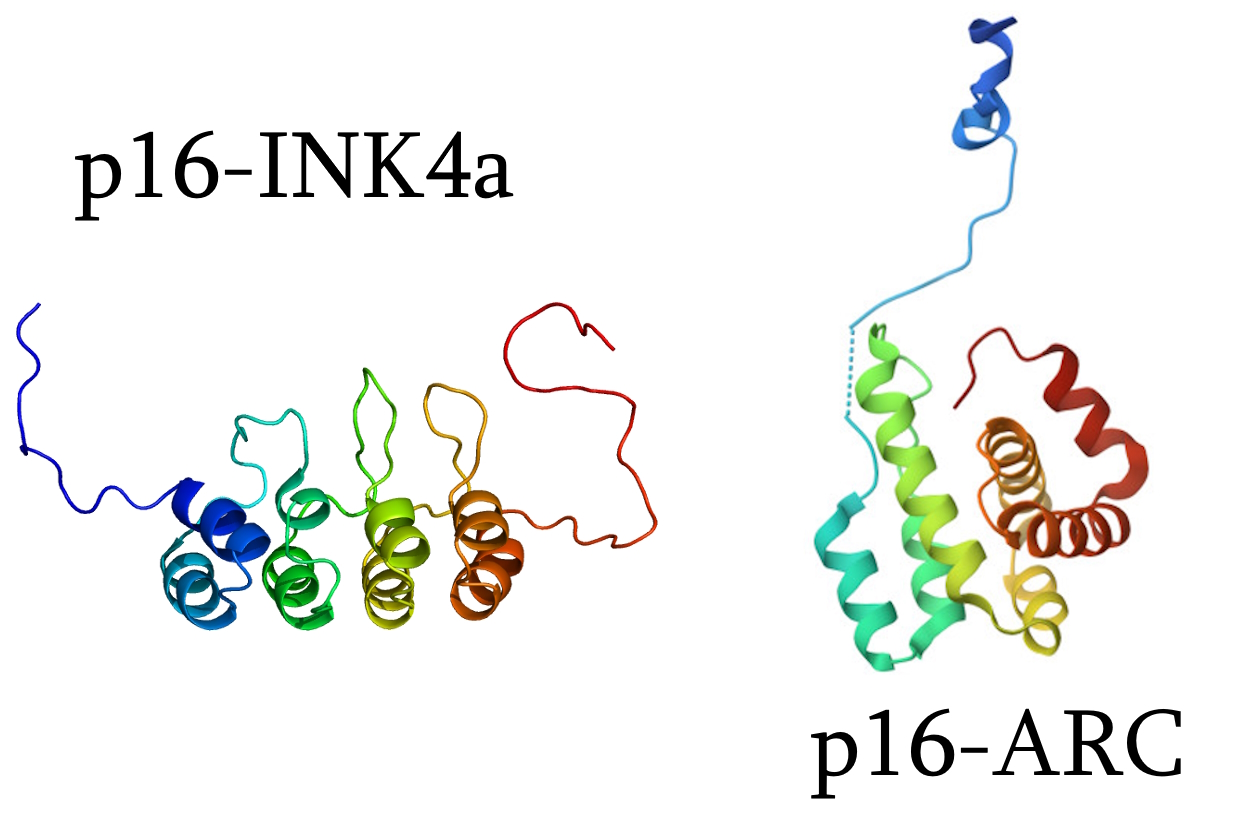
p16-INK4a is a protein encoded by the CDKN2A gene. It acts as a tumour suppressor by halting the cell cycle. Accumulation of p16-INK4a is said to trigger a state of “cellular senescence”. p16-INK4a is widely studied in cancer biology and is considered a key biomarker of ageing. Because p16-INK4a is frequently studied alongside related proteins (especially p21 and p53) it is often abbreviated simply as “p16”.
Actin-related protein 2/3 complex subunit 5 is a protein encoded by the ARPC5 gene. It is a comparatively less well-understood component of the actin cytoskeleton. Various synonyms are circulating including ARPC5, ARC16, dJ127C7.3, and, fatefully, p16-ARC (this is an alternative name listed by UniProt). Importantly, this protein is not structurally or functionally related to p16-INK4a, and is not known to be involved in cell cycle control or ageing.
So in this story we have two unrelated proteins; a critical tumour suppressor that I will continue to refer to as p16-INK4a, and a comparatively obscure component of the actin cytoskeleton, which I will refer to as p16-ARC.
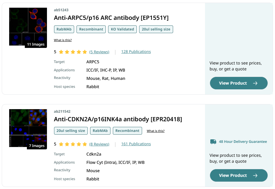
Antibodies are frequently used to investigate protein expression by western blot or immunostaining because of the specificity of their binding. Abcam is a major supplier of antibodies, and if you go to their website today and search for a “p16” specific antibody a list of potential choices is offered, importantly for this story a p16-ARC antibody is the first search result, the product code is “ab51243”. There is also another less frequently cited p16-ARC antibody “ab151303”. Similarly, if you head over to Santa Cruz (another supplier) you can find a p16-ARC antibody with the product code “sc-166760”. I think most people can guess where this is leading… How many researchers have muddled these two proteins and mistakenly ordered an antibody that binds p16-ARC when trying to investigate the expression of p16-INK4a?
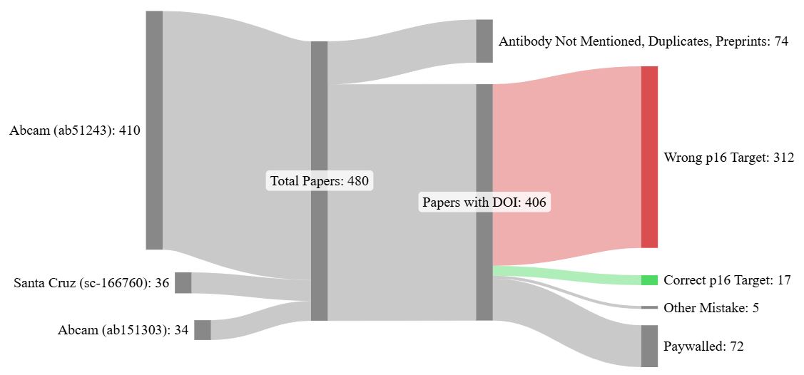
I decided to check, searching for the “ab51243”, “ab151303”, and “sc-166760” product codes in Google Scholar I found over 400 unique research papers mentioning at least one of these p16-ARC antibodies (after removing duplicates, false positives, preprints, and dead links). Of these papers I could access the full text of 334, with the remaining 72 articles being paywalled. I reviewed each accessible paper to determine whether the antibody used in the paper was correctly intended for p16-ARC or incorrectly used to try and bind p16-INK4a
Here is the astonishing result: 95% of these papers have got it wrong. The vast majority of researchers who purchased either ab51243, ab151303, or sc-166760 have tried to use these antibodies to investigate p16-INK4a expression! Only seventeen used these p16-ARC antibodies correctly. It is truly an astonishing unforced error which might sound unbelievable at first. You are invited to check my working, so here is a spreadsheet of the identified papers.
The implications are not good, to put it mildly. And these are not just insignificant papers. As I mentioned previously there are papers with hundreds of citations in high impact journals claiming to probe for p16-INK4a with antibodies which (I repeat) do not bind p16-INK4a! Well, don’t take it from me, it’s right there on Abcam’s product page:
“This antibody (ab51243) is specific to the p16 ARC protein […] There is no observed cross reactivity to the CDKN2A/p16INK4a protein.”
Let’s look at some examples, and we may as well start with the error as published by Nature. This paper comes from the lab of Jan Karlseder, CSO and Senior Vice President at Salk Institute in La Jolla, USA, his colleague Reuben Shaw (director of NCI Cancer Center at Salk), and their collaborators at University of Heidelberg in Germany. The paper is focused on the cell cycle, of which p16-INK4a is a critical regulator. Here we’ll look at extended data Figure 1d:
Joe Nassour, Robert Radford, Adriana Correia, Javier Miralles Fusté, Brigitte Schoell, Anna Jauch, Reuben J. Shaw, Jan Karlseder Autophagic cell death restricts chromosomal instability during replicative crisis Nature (2019) doi: 10.1038/s41586-019-0885-0
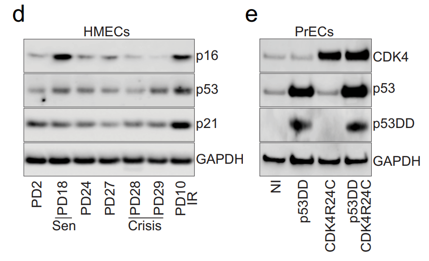
The western blot is labelled ambiguously as “p16” (although the figure also presents p53 and p21 blotting alongside p16 which is a major hint), in the methods section the antibody is listed as “P16 (Abcam ab51243)”. The figure is discussed in the main text, quote:
“Human mammary epithelial cells (HMECs) escape from senescence through spontaneous silencing of P16INK4A and enter crisis at PD27 (Extended Data Fig. 1c, d).”
So there is simply no question that the listed antibody is wrong as they have named the protein as p16-INK4a!
Karlseder eventually replied to out inquiry with:
“I will discuss this with the lead author and get back to you. We have worked with the correct p16 antibodies for years, so I would be surprised if the wrong one was used here.”

Capybara’s Adventures in Medicinal Chemistry
“Every now and again, it is a good idea to open the door of the clown car that is MD Anderson, and see who climbs out. Today is the turn of Kapil N. Bhalla. If you say his name quickly, it sounds a bit like “capybara”” – Sholto David
Next, here’s a figure published in Nature Medicine, this one from a team that is just down the road from me, led by Simon Leedham at the Wellcome Trust Centre for Human Genetics and the John Radcliffe Hospital (University of Oxford, UK). In this case the authors specify that they stained p16-INK4a in human samples, see below. This is wrong as they also used the p16-ARC antibody ab51243.
Hayley Davis, Shazia Irshad, Mukesh Bansal, Hannah Rafferty, Tatjana Boitsova, Chiara Bardella, Emma Jaeger, Annabelle Lewis, Luke Freeman-Mills, Francesc C Giner, Pedro Rodenas-Cuadrado, Sreelakshmi Mallappa, Susan Clark, Huw Thomas, Rosemary Jeffery, Richard Poulsom, Manuel Rodriguez-Justo, Marco Novelli, Runjan Chetty, Andrew Silver, Owen J Sansom, Florian R Greten, Lai Mun Wang, James E East, Ian Tomlinson, Simon J Leedham Aberrant epithelial GREM1 expression initiates colonic tumorigenesis from cells outside the stem cell niche Nature Medicine (2015) doi: 10.1038/nm.3750
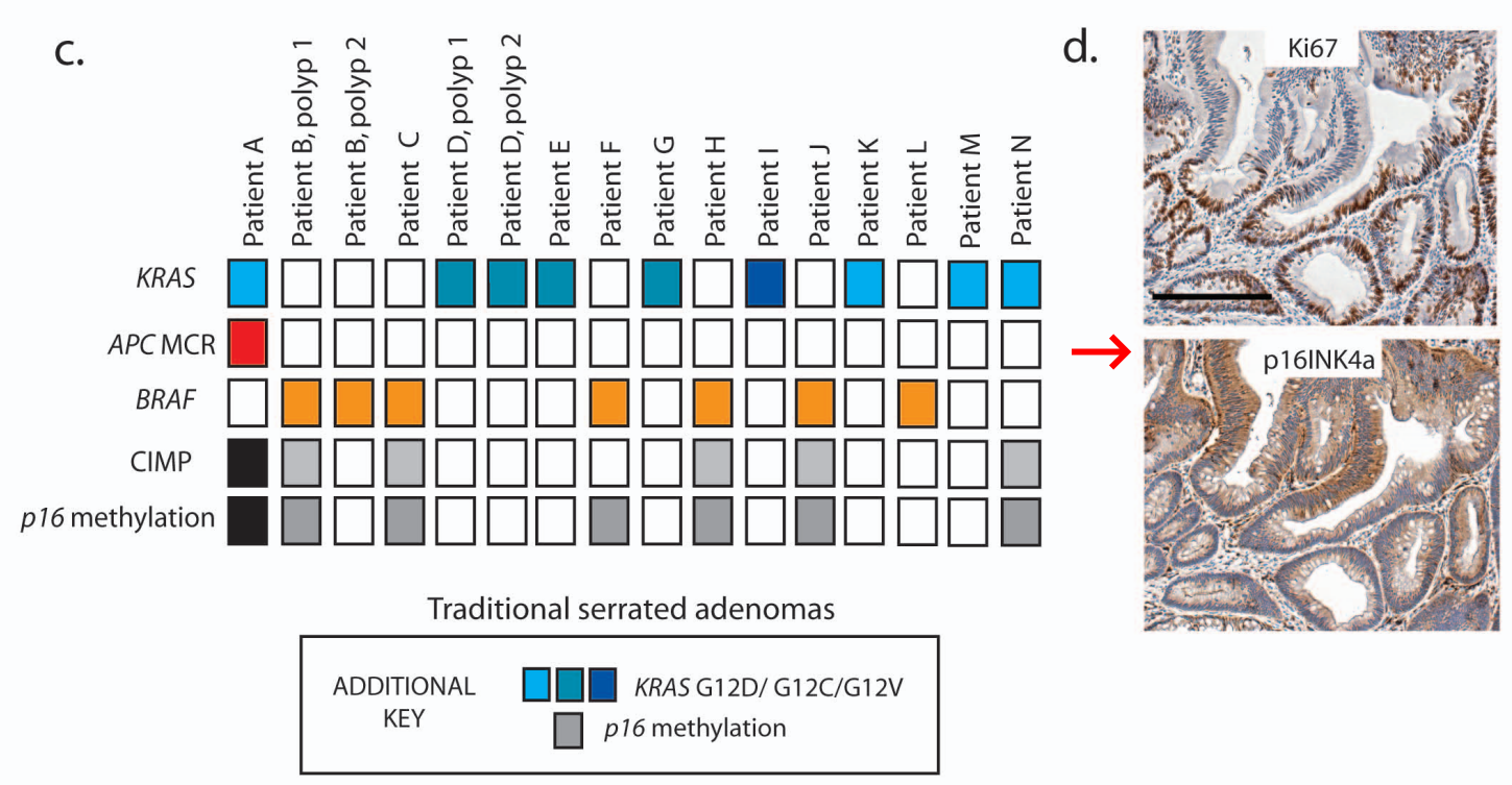
The authors, who include the Glasgow professor Owen Sansom (PubPeer record) and Michael Karin‘s former mentee Florian Greten (now eternally innocent professor at University of Frankfurt, Germany), mentioned another CDKN2A related antibody in this paper (PA1-30670 from ThermoFisher). They specified it as staining mouse p16-INK4a – this is also wrong though, PA1-30670 is supposed to bind p19-ARF which shares the same gene but is an entirely different protein because it is derived from a different reading frame (ARF stands for Alternative Reading Frame). So that’s awkward, they tried to stain p16-INK4a in humans and mice and failed both times by choosing the wrong antibody twice.

AACR conjures undead Count Fakula Michael Karin
What better distraction than the COVID-19 pandemic to revive one of the spookiest parasites in cancer research? AACR uses the COVID-19 cover to award Michael Karin, for his over 50-paper-strong record of data fakery.
It would be unfair to pick on Oxford, so here’s a Cambridge team, led by emeritus professor Graham Burton, trying to western blot human placenta samples for p16-INK4a with the same wrong p16-ARC antibody (ab51243). Burton used to be founding chair of the Cambridge Reproduction initiative, allow us the pun to ask how reproducible his p16 result is:
Tereza Cindrova-Davies, Norah M.E. Fogarty, Carolyn J.P. Jones, John Kingdom, Graham J. Burton Evidence of oxidative stress-induced senescence in mature, post-mature and pathological human placentas Placenta (2018) doi: 10.1016/j.placenta.2018.06.307
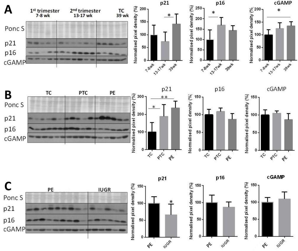
Interestingly, the methods state that they also performed immunohistochemistry with this antibody although they only seem to show the results for p21 staining… I wonder why?
Here’s another example, this time from Immunity published by Cell Press, this team is from Stanford University School of Medicine, led by Jörg Goronzy and Staford’s former centre director Cornelia Weyand, both are now at Mayo Clinic in Rochester. Again, this paper also uses the ambiguous name “p16” but it is clear from the text that the authors intended to investigate p16-INK4a and not p16-ARC, for example, quote (highlight mine):
“To better define the aging process in naive CD4+ CD45RA+ T cells, we assessed the aging-associated cell-cycle inhibitors p16, p21, and p53″
Only p16-INK4a is an aging associated cell-cycle inhibitor related to p21 and p53, p16-ARC is certainly not. Now here’s Figure 5E and F:
Yinyin Li, Yi Shen, Philipp Hohensinner, Jihang Ju, Zhenke Wen, Stuart B. Goodman, Hui Zhang, Jörg J. Goronzy, Cornelia M. Weyand Deficient Activity of the Nuclease MRE11A Induces T Cell Aging and Promotes Arthritogenic Effector Functions in Patients with Rheumatoid Arthritis Immunity (2016) doi: 10.1016/j.immuni.2016.09.013
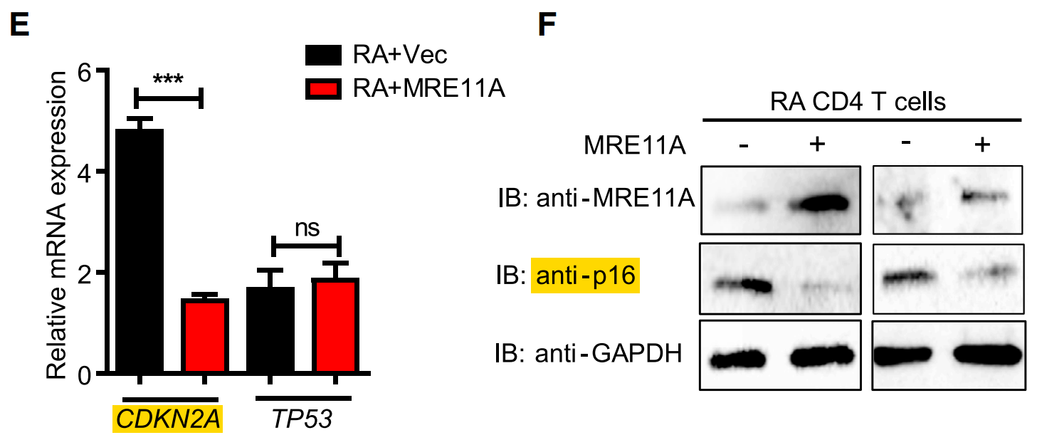
Note that CDKN2A is the gene for p16-INK4a making it clear (if it wasn’t already) which p16 the authors wished to investigate. We can also look at Figure 6H, and it is helpful to share the discussion of this figure, again linking the CDKN2A gene to the p16 protein (highlights mine):
“To examine a possible link between tissue invasiveness, inflammatory capability and cellular aging, we quantified tissue expression of CDKN2A, CDKN1A, and TP53 transcripts and measured p16 protein expression by dual-color immunohistochemistry.”
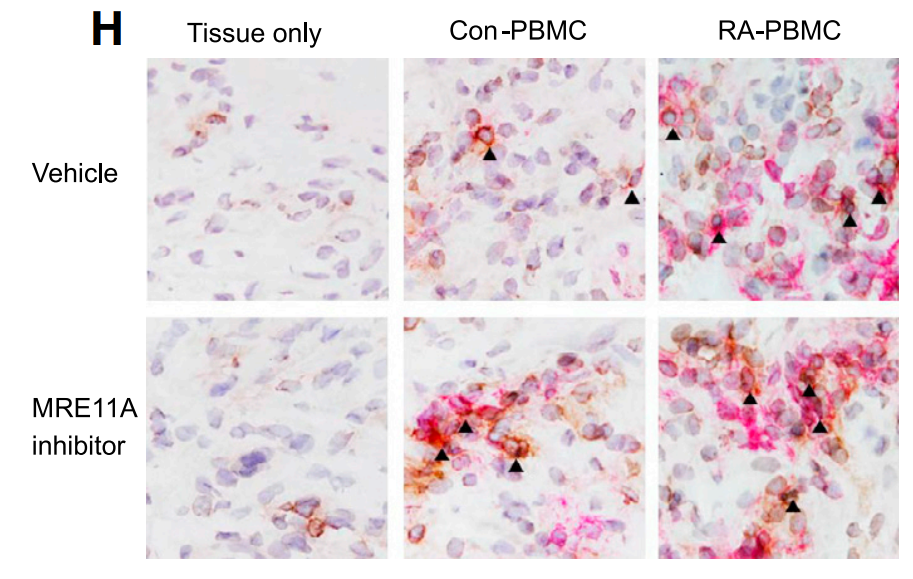
Here is a quote from the corresponding methods section (highlights mine):
“Slides were incubated with a primary antibody cocktail overnight at 2C to 8C. The cocktail contained both monoclonal mouse anti-human CD3 (Clone F7.2.38; 1:100, Dako), and anti-p16 ARC antibody (EP1551Y; 1:100, Abcam, ab51243).”
Interestingly, here the authors correctly describe the antibody as a p16-ARC antibody (presumably copied directly from the Abcam product page) but it is quite clear they had no interest in the actin related protein… the word “actin” does not even appear in this paper!

Scientific Reports 2025: A Year in Review
“In this blog I write about papers published by Scientific Reports in 2025, so we could consider it to be a sort of “wrap-up” of highlights and special achievements in the world’s biggest scientific journal™ in 2025.” – Sholto David
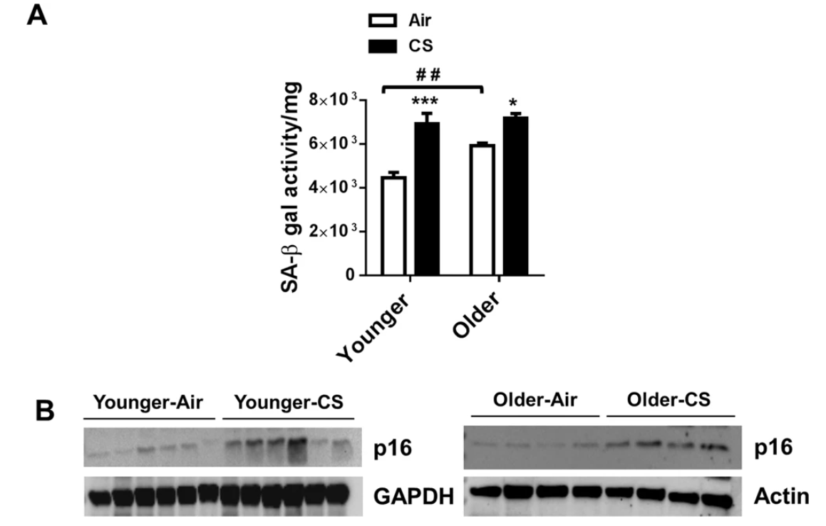
This isn’t the only paper from Rochester academics. And I was not going to write a blog without poking fun at Scientific Reports and their “excellent team”! So here is a study led by Irfan Rahman on a mouse model of COPD which they produce by forcing the mice to smoke “research grade” cigarettes. And then they blotted the wrong p16 (ab51243 again).
Kahkashan Rashid, Isaac K. Sundar, Janice Gerloff, Dongmei Li, Irfan Rahman Lung cellular senescence is independent of aging in a mouse model of COPD/emphysema Scientific Reports (2018) doi: 10.1038/s41598-018-27209-3
You might be interested to learn that University of Rochester’s professor Rahman is not only among the “top 0.03% of Research Scholars in the world“, but also owner of a disastrous PubPeer record, which, what a surprise, includes many duplicated western blots and microscopy images. Also on the topic of smoking, see for example Moodle et al 2004, Marwick et al 2006, Marwick et al 2010, Edirisinghe et al 2008, Arunachalam et al 2010 or Yogeswaran et al 2022, or if you are rather into curcumin, see Meha et al 2008. No prizes for guessing how Professor Rehman made the wrong p16 antibody produce the right results in his lab.

Lashing out at Toxicology Reports
“What exactly will Lash and Elsevier do with these 115 problematic papers? I can only expect a painfully inadequate response.” – Sholto David
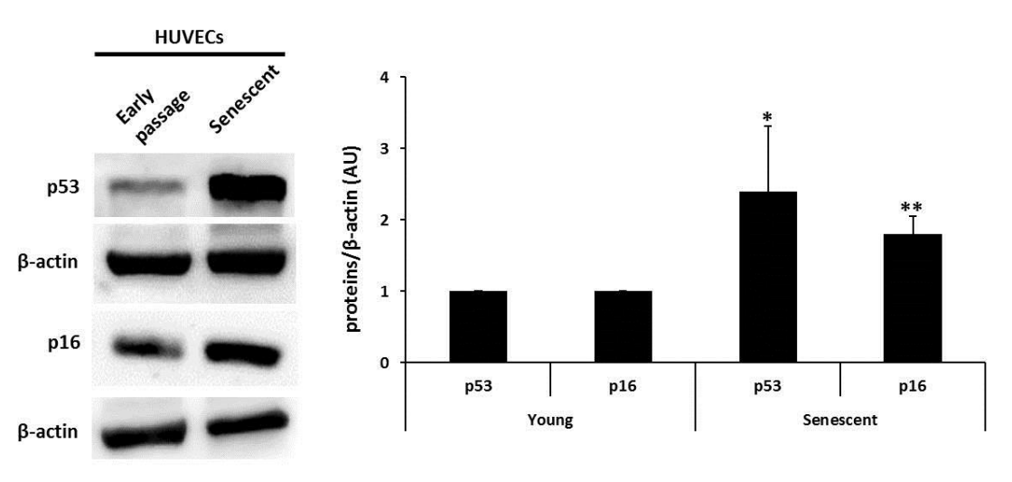
What’s better than Scientific Reports? MicroRNA + Scientific Reports. A legendary partnership. Here the Spanish authors from Madrid describe p16 as a “conventional cellular marker of senescence” and western blot with ab51243 again.
Matilde Alique, Guillermo Bodega, Chiara Giannarelli, Julia Carracedo, Rafael Ramírez MicroRNA-126 regulates Hypoxia-Inducible Factor-1α which inhibited migration, proliferation, and angiogenesis in replicative endothelial senescence Scientific Reports (2019) doi: 10.1038/s41598-019-43689-3
Sorry, back to serious research in proper journals and this next paper is from Cancer Cell published by Stephen Baylin at The Sidney Kimmel Comprehensive Cancer Center at Johns Hopkins University in USA. Once again we’re looking at a p16 western blot which was performed with the ab51243 antibody, the authors describe p16 here as a “known anti proliferative gene” (i.e. a tumour suppressor) meaning they must have intended to target p16-INK4a.
Xiangqian Kong, Jie Chen, Wenbing Xie, Stephen M. Brown, Yi Cai, Kaichun Wu, Daiming Fan, Yongzhan Nie, Srinivasan Yegnasubramanian, Rochelle L. Tiedemann, Yong Tao, Ray-Whay Chiu Yen, Michael J. Topper, Cynthia A. Zahnow, Hariharan Easwaran, Scott B. Rothbart, Limin Xia, Stephen B. Baylin Defining UHRF1 Domains that Support Maintenance of Human Colon Cancer DNA Methylation and Oncogenic Properties Cancer Cell (2019) doi: 10.1016/j.ccell.2019.03.003
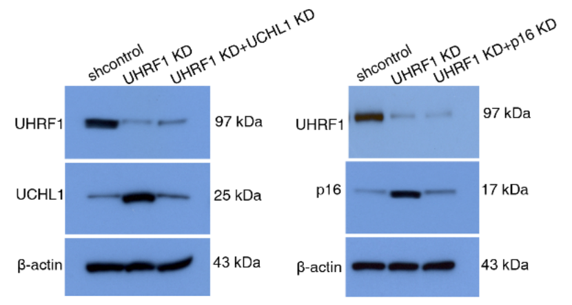
In fact, Baylin and his former mentee Limin Xia are repeat offenders because they published another western blot using the same wrong antibody also in Cancer Cell two years prior! In this case they also discussed p16 in the context of senescence confirming their intended target.
Limin Xia, Wenjie Huang, Marina Bellani, Michael M. Seidman, Kaichun Wu, Daiming Fan, Yongzhan Nie, Yi Cai, Yang W. Zhang, Li-Rong Yu, Huili Li, Cynthia A. Zahnow, Wenbing Xie, Ray-Whay Chiu Yen, Feyruz V. Rassool, Stephen B. Baylin CHD4 Has Oncogenic Functions in Initiating and Maintaining Epigenetic Suppression of Multiple Tumor Suppressor Genes Cancer Cell (2017) doi: 10.1016/j.ccell.2017.04.005
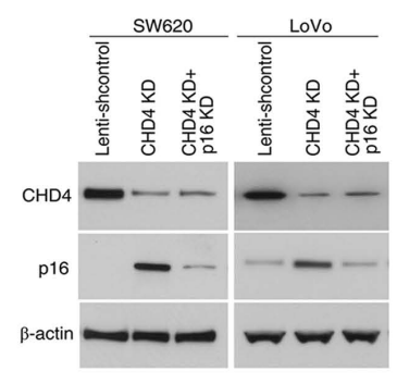
Xia is now professor at Huazhong University of Science and Technology in Wuhan. And as for clues how she made this wrong antibody work: also Xia has her own PubPeer record of duplicated and cropped microscopy images, see for example Li et al 2018 or Huang et al 2015, the latter somehow corrected in 2022.

Perfect Blots
“A coordinated review of the cited publications and underlying data is currently underway” – Hiroki Kuniyasu
Here’s a recent paper in Science Advances from Jonathan Keller’s lab at the NIH’s National Cancer Institute in Bethesda, USA. From the abstract regarding the analysis of the results: “Mechanistically, p16 expression, senescence, and apoptosis were increased…” making it clear which p16 they were interested in. Here is Figure 5:
Shweta Singh, Kristbjorn O. Gudmundsson, Tanmoy Sarkar, Brad L. Jakubison, Holly M. Morris, Sandra Burkett, Karim Baktiar, Gary T. Pauly, Dina M. Sigano, Joel P. Schneider, Lino Tessarollo, Jonathan R. Keller Id1 promotes clonal hematopoiesis in mice with Tet2 loss of function Science Advances (2025) doi: 10.1126/sciadv.adr5867
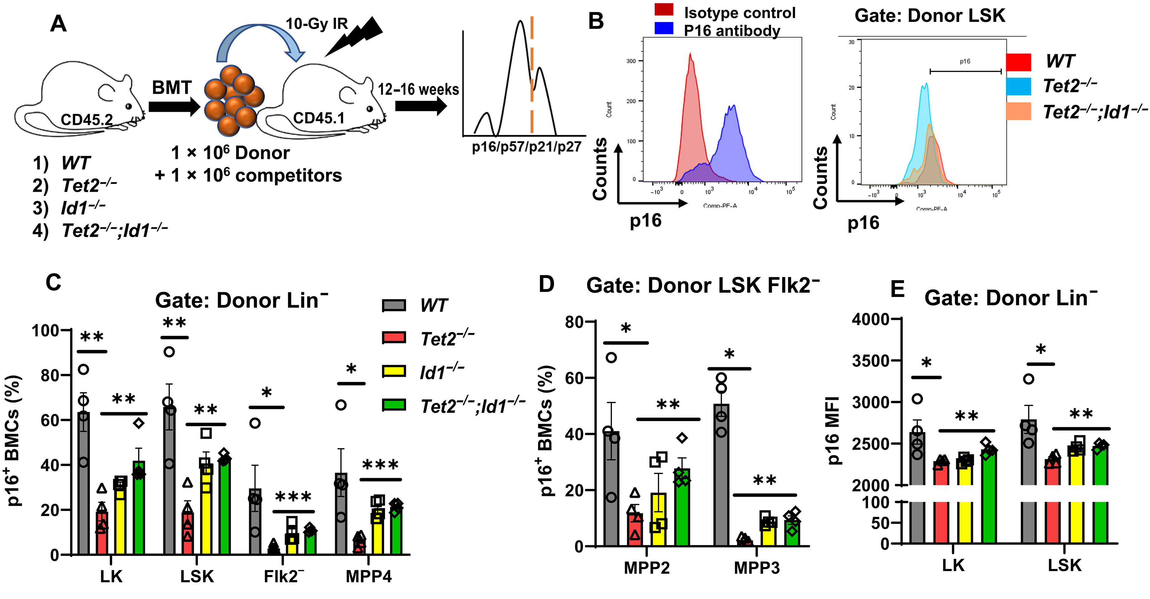
I don’t think it’s quite worth trying to explain the whole figure but the method requires an antibody to stain cells as p16 positive or negative and the only p16 antibody provided in the methods is sc-166760, a p16-ARC antibody. In fact, Keller’s lab has been using this wrong antibody since at least 2018 (seven years earlier) to try to stain p16-INK4a, as he has another publication in the Cell family journal Stem Cell (Singh et al 2018). Keller has now graduated to NCI Scientist Emeritus.
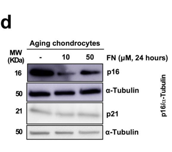
How about a Russian-American-Spanish anti-ageing collaboration on the subject of senolytic drugs? If the Spanish succeed in reversing ageing they might reach their long-held ambition of 100% youth unemployment. Alas – they tried to blot p16-INK4a but used the wrong antibody (ab51243) and the experiments are doomed. I’m joking of course, senolytics isn’t real research anyway, it doesn’t matter what you blot.
Uxía Nogueira-Recalde, Irene Lorenzo-Gómez, Francisco J. Blanco, María I. Loza, Diego Grassi, Valery Shirinsky, Ivan Shirinsky, Martin Lotz, Paul D. Robbins, Eduardo Domínguez, Beatriz Caramés Fibrates as drugs with senolytic and autophagic activity for osteoarthritis therapy eBioMedicine (2019) doi: 10.1016/j.ebiom.2019.06.049
Here’s another US-based lab blotting the wrong p16. The lead author Victor Thannickal is now Chair of the John W. Deming Department of Medicine at Tulane University. Again these authors used the ab51243 antibody.
Yan Y. Sanders, Hui Liu, Xiangyu Zhang, Louise Hecker, Karen Bernard, Leena Desai, Gang Liu, Victor J. Thannickal Histone Modifications in Senescence-Associated Resistance to Apoptosis by Oxidative Stress Redox Biology (2013) doi: 10.1016/j.redox.2012.11.004
Also Professor Thannikal has a PubPeer record of questionable science, with duplicated western blot bands and microscopy images, see for example Jiang et al 2021, Ghatalk et al 2017, Huang et al 2011 or Hecker et al 2009.
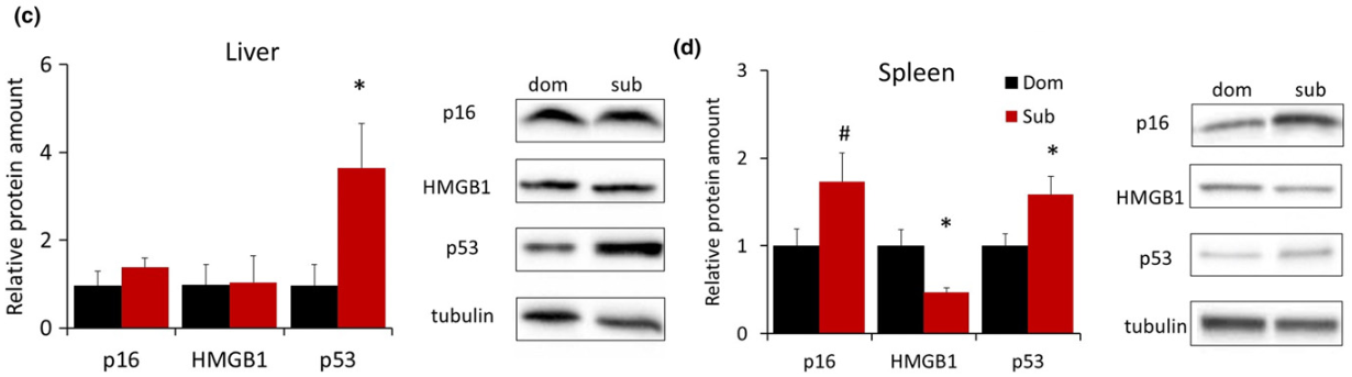
The following is a charming study that seems to involve bullying mice to death; title “Social stress shortens lifespan in mice”. The authors, mostly at the University of Minnesota, claimed to measure a senescence signal in mouse organs, partly by p16-INK4a expression… but they’ve used the wrong antibody (ab51243).
Maria Razzoli, Kewir Nyuyki-Dufe, Allison Gurney, Connor Erickson, Jacob McCallum, Nicholas Spielman, Marta Marzullo, Jessica Patricelli, Morito Kurata, Emily A. Pope, Chadi Touma, Rupert Palme, David A. Largaespada, David B. Allison, Alessandro Bartolomucci Social stress shortens lifespan in mice Aging Cell (2018) doi: 10.1111/acel.12778
The coauthor and Minnesota professor David Largaespada is a former collaborator of the infamous Catherine Verfaillie, and coauthor on her now-retracted Nature paper Jiang et al 2002, which fraudulently claimed that bone marrow cells were pluripotent.

Catherine Verfaillie, the Zombie Scientist of KU Leuven
Catherine Verfaillie is a zombie scientist: her past stem cell research long discredited, but she still is an influential and very well funded star of Belgian science. Now Elisabeth Bik had a fresh new look at Verfaillie’s papers
Another coauthor, former Indiana University dean David Allison (now at University of Alabama at Birmingham), is actually a leading champion of reproducibility, celebrated by Nature:
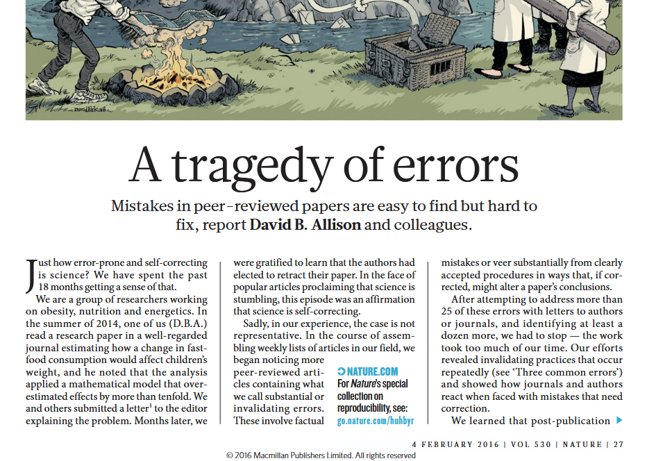
We will see how hard it will be to fix this mistake in Allisson’s own paper.
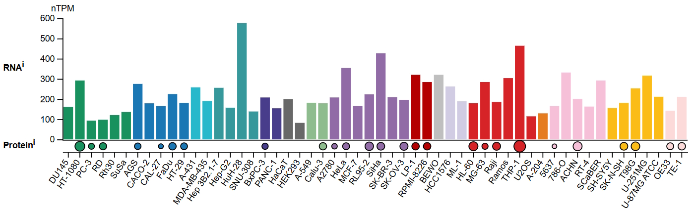
A natural question that follows from these observations: If p16-ARC has no relationship to the cell cycle or ageing, what exactly should we see when it is accidentally investigated in these papers? Data collated by the Human Protein Atlas for p16-ARC (ARPC5) shows comparatively high levels of transcription and (where measured) protein expression across commonly used cell lines. p16-ARC is known to be a component of the actin cytoskeleton, so logically (in most circumstances) it should track β-actin expression.
In other words, as far as western blots go, it should look like a loading control!
In fact several papers appear to (accidentally) stumble into this finding. Here’s an example; researchers from Fudan University in China analysed the response of senescence related genes in cells exposed to increasing doses of radiation:
Jiangtao Bai, Yuyang Wang, Jianping Wang, Jianglong Zhai, Feilong He, Guoying Zhu Irradiation-induced senescence of bone marrow mesenchymal stem cells aggravates osteogenic differentiation dysfunction via paracrine signaling American journal of physiology. Cell physiology (2020) doi: 10.1152/ajpcell.00520.2019
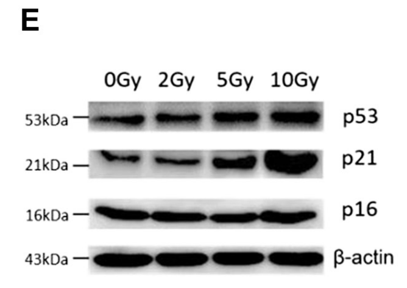
Figure 4E: expression of p53, p21 and p16 was measured by quantitative RT-PCR and Western blot analyses.
Quote from this paper related to the above figure (highlight mine):
“Furthermore, results of the mRNA and protein levels of senescence-related genes showed that irradiation caused upregulation of p53 and p21 from 2 Gy to 10 Gy, whereas p16 had no obvious change at any dose of gamma rays (Fig. 4, E–G)”
The explanation, of course, is that they were not blotting p16-INK4a, but p16-ARC, which will likely track β-actin expression (as it does here). Another paper with a similar experiment made the same observation, this time from Third Military Medical University in Chongqing:
Lili Chen, Qian Ran, Yang Xiang, Lixin Xiang, Li Chen, Fengjie Li, Jiang Wu, Chun Wu, Zhongjun Li Co-Activation of PKC-δ by CRIF1 Modulates Oxidative Stress in Bone Marrow Multipotent Mesenchymal Stromal Cells after Irradiation by Phosphorylating NRF2 Ser40 Theranostics (2017) doi: 10.7150/thno.17853
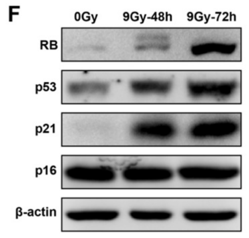
Figure 1. (f) Proteins (RB, p53, p21, and p16) related to cell senescence were detected by western blot after irradiation at 48 and 72 h. The representative immunoblots (left panel) and relative levels of proteins normalized to β-actin (right panel) are shown.
Quote from the authors regarding this figure (highlight mine):
“Several proteins related to cell senescence were detected by western blot after irradiation at 48 and 72 h (Figure 1F). The results showed that protein levels of RB, p53, and p21 significantly increased, confirming that cell senescence was induced by irradiation, while no remarkable change of p16 was observed.”
Well, once again, the explanation is rather simple, the expression of p16-ARC should probably track β-actin (and it does!).

Luck in Sight
“It seems hard to accept any explanation that doesn’t somehow incriminate most of the people involved” – Sholto David
Perhaps more interesting is that so many of these papers have managed to produce the “expected” behaviour of p16-INK4a in response to various experimental manipulations despite using the wrong antibody. Let’s look at another example, in eLife, the authors affiliated in China and Australia measured what they believed to be p16-INK4a expression by immunofluorescence and western blot, quote:
“p16 expression decreased after siRNA-mediated knockdown, which decreased the proportion of NP cells that demonstrated a senescent phenotype. By contrast, p16 overexpression caused a marked opposite effect…”
Hui Che, Jie Li, You Li, Cheng Ma, Huan Liu, Jingyi Qin, Jianghui Dong, Zhen Zhang, Cory J Xian, Dengshun Miao, Liping Wang, Yongxin Ren p16 deficiency attenuates intervertebral disc degeneration by adjusting oxidative stress and nucleus pulposus cell cycle eLife (2020) doi: 10.7554/elife.52570
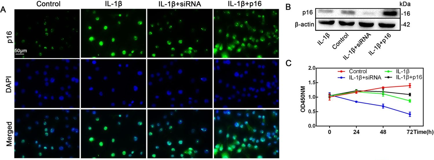
The authors only provided one p16 antibody product code (ab51243), along with specified dilutions for western blotting and immunofluorescence.
What are we to make of cases like this where the wrong antibody was used but the authors still manage to rustle up interpretable results? One explanation is that the data has been manipulated to conform to the researcher’s expectations or desires… i.e. fraud. And some of these are the most obvious frauds, at least four have been retracted, here’s one in the style of Elisabeth Bik’s famous tadpole papermill. It wasn’t noted at the time, but they used the wrong antibody for p16-INK4a here (ab151303 is a lesser cited p16-ARC antibody).
Zhijia Yang, Guoqing Li, Chao Ding, Wencong Sun, Ji Zhang Long non-coding RNA HULC exerts oncogenic activity on papillary thyroid cancer in vitro and in vivo Artificial Cells Nanomedicine and Biotechnology (2020) doi: 10.1080/21691401.2019.1703730
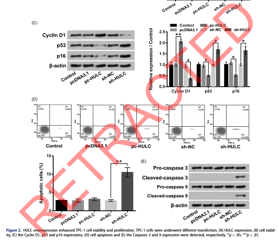
Retraction September 2025: “In 2021, concerns were identified regarding image integrity of the article, and an expression of concern was published […] In 2025, additional image integrity concerns were identified by the publisher in western blot Figures 2C, 2E, 3C, 3D, 5C, 6C, 6D, 7A, 7B, flow cytometry Figure 2D, and qRT-PCR Methodology.”
Another potential explanation for these impossible results is that the experiments actually were performed with a p16-INK4a specific antibody, but at the moment of writing the paper, a p16-ARC antibody code was retrieved from Abcam or Santa Cruz’s website and inserted into the methods section erroneously. This second explanation might seem more charitable in some sense, but I’m not sure it is… are we to believe that antibody codes in papers are selected at random by a lab tech based on the first search result? For papers to be meaningful the methods have to describe what was actually done and the results have to show what was observed.

The Men Who Stare At Mice
“Do Cohen’s colleagues and superiors know or care that he hosts wizards in his lab? Or perhaps this is simply common place, wizards roam throughout MD Anderson free range, blasting the cancer-mice with their mind powers.” – Sholto David
In the end there is likely a mixture of different explanations ranging from outright fabrication, selective reporting, writing errors, and some teams blindly publishing contradictory findings without further questioning or curiosity. There is something unsettling about the idea that hundreds of papers can be published using completely the wrong antibody without anyone noticing, as if the research results are only loosely connected with biological reality. How many antibodies could you swap for loading controls until someone actually takes notice of what the results are telling them?
Anyway, I’m not going to write about all the papers here, but I will try to post them all to PubPeer eventually. There are plenty more interesting examples, including one paper where some Chinese researchers blasted some cells into space (Mao et al 2024), then stained them with the wrong p16 antibody, shame, perhaps they’ll have to repeat the rocket launch? Or what about this experiment on microplastics and mouse testicle ageing (Wu et al 2023), already cited over 100 times I expect it will be extensively laundered into the scientific literature by very serious reviews, but how competent are the researchers who can’t even order the right antibody?

Cell Cycle of Angry Axe-Wielding Tribbles
“For the most competitive papers, an ultra-rapid review (by members of the Editorial Board) is necessary to publish them a few days after submission.”, Misha Blagosklonny, on how his journals became papermill fraud bonanza
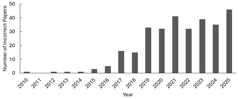
I did wonder how widespread this problem is. Well, speculation is irresponsible and unscientific so let’s do that. If we take this question to mean something like: “Of those papers that probe for p16-INK4a, what percentage mistakenly used a p16-ARC antibody?” I would guess likely less than 10% but maybe more than 5%. If that sounds like a lot… Well, I already have a list of over 300 incorrect papers so in order to prove me wrong you would need to find at least 6000 papers which correctly used a p16-INK4a antibody. Based on my searches for valid p16-INK4a antibody product codes I think that might be difficult! And this remains a highly active problem, in fact 2025 was a record year for wrong p16 antibody papers. As long as the first search result for “p16” on the Abcam website is a p16-ARC antibody I think this will continue.
For sure this error does not impact as many papers as related incidents where non-specific antibodies have been distributed by manufacturers, Entrop et al convincingly argued in 2024 that >1400 papers have used a non-specific antibody for BAX sold by Santa Cruz (sc-7480). But the case presented here has a different quality, more like an own goal, given that these p16-ARC antibodies were never claimed to be p16-INK4a antibodies in the first place.
So here is a quick post mortem on what went wrong here: On the side of the researchers who made this error, obviously the naming collision is unfortunate, and these proteins have similar names precisely because they are about the same molecular weight, thus making the confusion harder to spot by western blot because they will run to the same size on a gel.

Shocked, angered and appalled
“I have been following the comments on PubPeer, and have been shocked, angered and appalled by the issues […] there can be no explanation for this other than systemic fraud “- Prof Gareth Williams, UCL
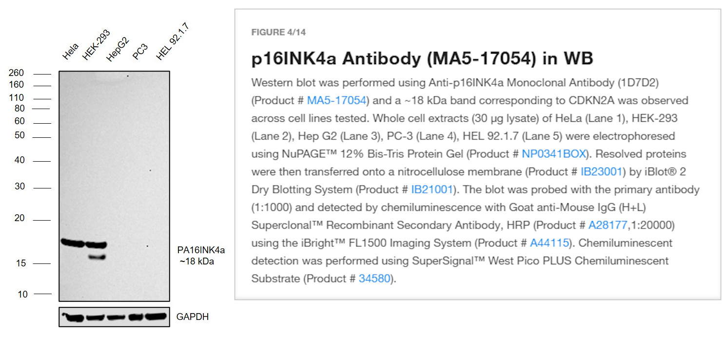
On the side of not making thoroughly clownish mistakes this could have been so easily been avoided by running simple well-established controls. ThermoFisher has a very clean example for what this might look like on one of their p16-INK4a antibody pages. Well, maybe don’t trust a ThermoFisher blot (see May 2026 Shorts as to why), but in principle they show a sensible verification that would have immediately caught this problem, the key control here is the PC-3 cell line (presumably the lung carcinoma cell line) which has a homozygous CDKN2A deletion and therefore cannot express p16-INK4a. Not to be confused with the prostate cancer cell line with the same name, but I digress, another naming confusion for another day. Similar experiments with knockouts or siRNA are possible.
However, it seems as if most researchers purchased these p16-ARC antibodies and must have immediately implemented them in their research without further testing or controls. The Nature paper cited above includes a statement about antibody validation in the reporting summary: “Antibodies were validated by the size of the band and by targeting the corresponding product by siRNA.” – it would be interesting to see the data!
I’m quite sure there are more soundalike/sizealike proteins to find. Amusingly the senescence related protein usually referred to as “p21” also has an imposter p21-ARC protein! In these two papers by Chinese researchers based in Chengdu the team have managed to simultaneously blot for the wrong p16 (sc-166760) and the wrong p21 (sc-166630).
Peng Zhang, Qian Wang, Lulingxiao Nie, Rui Zhu, Xinyi Zhou, Pengfei Zhao, Ning Ji, Xing Liang, Yi Ding, Quan Yuan, Qi Wang Hyperglycemia-induced inflamm-aging accelerates gingival senescence via NLRC4 phosphorylation Journal of Biological Chemistry (2019) doi: 10.1074/jbc.ra119.010648
The second paper was published in the previously well regarded Journal of Biological Chemistry:
Lulingxiao Nie, PengFei Zhao, Ziqi Yue, Peng Zhang, Ning Ji, Qianming Chen, Qi Wang Diabetes induces macrophage dysfunction through cytoplasmic dsDNA/AIM2 associated pyroptosis Journal of Leukocyte Biology (2021) doi: 10.1002/jlb.3ma0321-745r
This p21 error not only effects Chinese labs, but even the highly respectable former rector of the University of Messina, one Salvatore Cuzzocrea, who is surely familiar to readers.

Cuzzocrea’s Magnificent Fall
“These unscrupulous charlatans in Messina should be fired on the spot tomorrow morning, forced to return twenty years of undeserved wages and sent to work the land” – Aneurus Inconstans
So, here it is:
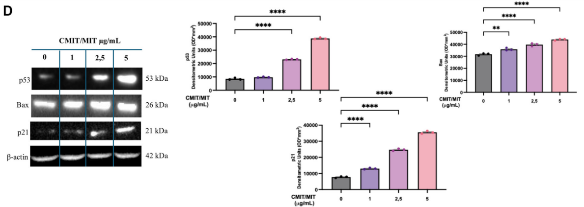
Francesco Molinari, Nicla Tranchida, Francesca Inferrera, Roberta Fusco, Caterina Faggio, Federica Impellitteri, Salvatore Cuzzocrea, Marika Cordaro, Rosanna Di Paola Biocide mixture (CMIT/MIT) induces neurotoxicity through the upregulation of the MAPKs signaling pathways Journal of Neurophysiology (2025) doi: 10.1152/jn.00104.2025
And I could also talk about p38 and direct you to Han et al 2023… but I must stop writing now, or this could go on forever.
I considered whether I should email Abcam and ask them to change the name of the ab51243 antibody on their website to reduce confusion, but I won’t be doing that. It feels too much like asking Abcam to put the bowling lane bumpers up. I will instead continue to watch the carnage, perhaps it has only just started!

Donate!
If you are interested to support my work, you can leave here a small tip of $5. Or several of small tips, just increase the amount as you like (2x=€10; 5x=€25). Your generous patronage of my journalism will be most appreciated!
€5.00


Fascinating. A few comments
LikeLike
If journals were more insistent, the authors would have quietly replaced the wrong antibody’s code with the right one, and we would never know about the fraud which clearly happened to get those results right.
LikeLike
“There is likely a wealth of useful information in these papers, since they have looked at cytoskeletal responses – the wily will dig through these 400+ papers, design some experiments and make discoveries.”
How do you know? Just as reliable as papers, where mistakes are not made? There are already more papers than anybody can read, why dig through these 400+?
“It would help if there was an insistence by journals to use the systematic UniProt name in all instances with the old name(s) in parentheses at first use.”
I see, it’s the journals’ fault.
I agree with Leonid, checklists are checklists for the careless at the highest.
LikeLike
Maybe UniProt AC/ID would be even better (we usually use them, at least in supmat, but should be in main text). Then even if the canonical name changes, the identifier remains.
LikeLike
The recommendation from UniProt is to use Accession numbers (https://www.uniprot.org/help/accession_numbers ).
LikeLike
‘… whether I should email Abcam and ask them to change the name of the ab51243 antibody on their website to reduce confusion.’
Hmm… they probably copied the ‘order number’ from other papers. It might even be a ‘self-propagating’ plagiarism.
LikeLike
Yes, but they also ordered this same antibody!
Imagine the scene. Several PhDs and postdocs fail to produce the desired p16INK result with that wrong antibody, get punished by PI. But one bright student succeeds where others fail. He or she publishes beautiful papers in big journals and gets beautiful letters of recommendation.
LikeLike
But then, a few years later, the PubPeer comments start to appear……
LikeLiked by 1 person
After some additional thought (as I mentioned to Leonid before) I have decided the best solution is in fact to ask Abcam to change the antibody description to explain that it does bind p16-INK4a after all. This will save a lot of trouble for all these authors and there now hundreds of papers to support this reassignment.
LikeLiked by 2 people
‘Yes, but they also ordered this same antibody!’ How to be sure? Unless the invoices are included in the supplemental material, I for one, suspect that there was nothing ordered at all, just like there were no tests done, everything is fake.
LikeLike
“In other words, as far as western blots go, it should look like a loading control!” As it happens, ARPC5 is one of my favorite loading controls. As it also happens, the antibody we use, ab118459 has also been mistaken for an anti-CDKN2A antibody in several papers. Indeed this has become such a SNAFU that the NIH-funded clown car has corrupted Google AI – look it up for yourself!
LikeLiked by 1 person
I think it is expected that LLMs will be hoodwinked by these problems. At least one person on twitter seemed to think “AI” could be used to search for this type of error. I’m less than convinced. Regular expressions would work fine. Interesting, thanks for sharing about ARPC5. Another antibody to add to the list.
LikeLike
Actually, the query needed is “antibody ab118459”.
Link to the antibody datasheet is here: https://www.abcam.com/en-us/products/unavailable/arpc5p16-arc-antibody-ab118459
LikeLiked by 1 person
Just for S&G, I told Google they’re wrong. Watching for the kill drones…..
LikeLike
Wow, they have already fixed it. All hail our cyber-overlords.
I can provide a screenshot of the previous AI summary if anyone wants to see it.
LikeLike
Aaaaaand now they’ve fucked it up again by resurrecting all the false/fake shit. I have learned a lot about Google AI today. Too damn democratic.
LikeLike
A minor correction: David Allison is currently at Baylor University. (He was at the University of Alabama at Birmingham before he was at Indiana University. He went from Indiana to Baylor.)
LikeLike
Thanks for the info!
But admit it, his Nature piece on reproducibility gets a very new and hilarious meaning now, no?
LikeLike
Not sure why my reply to you came out as a reply to owlbert, and I don’t see how to delete it. So, in the proper place, with a small clarification:
I agree that there’s an irony, but it’s not the first such irony in science. Probably not the last–unfortunately. (I know David, and I’d expect he’ll be as distressed about the potential problem with the antibodies as you are or I am. I doubt he handled that aspect of the research, for whatever it’s worth, but all of the authors have to deal with the problem.)
LikeLike
if it’s supposed to behave like a loading control but in many cases shown here clearly doesn’t, I’d prefer the more parsimonious and more generous explanation that the methods are wrong but not the antibody (e.g., some researcher used an antibody already in the fridge and only wrongly looked up the number when writing the methods, or that antibody-ordering and methods-writing happened multiple years apart). still not ideal in terms of reproducibility, but far less problematic, and doesn’t feed the narrative that all this is fake.
LikeLike
I don’t know if it is more generous? After all, writing down what you actually did is the easy part… As for being more parsimonious? Depends on how reliable you think an average biology paper is…
LikeLike
The most parsimonious interpretation is that they all used the p16-ARC/ARPC5 antibody stated, and it did serve as a loading control since that protein is expressed abundantly by all cells. Thus a blot where levels vary indicates unequal protein loading, simple as that. Of course as for the interpretations, well…. cancer researchers, eh?
LikeLiked by 1 person
I happened to be privy to an email exchange where authors admitted to have indeed used that wrong antibody, but assured that the conclusions remain unaffected.
Parse this.
LikeLiked by 1 person
Wow, shocking findings! I looked up purchasing out of curiosity, and found that even some vendors appear to have mixed them up! Santa Cruz Biotechnology calls it a tumor suppressant in the description of the product, with no mention of its real function! So it’s possible people have been purchasing it believing they have the right antibody. Other, better suppliers don’t seem to have made this mistake, but it’s still shocking.
Then again, I suppose you could say there’s a body of research showing that it is a tumor suppressant…
LikeLiked by 1 person
Ha! That’s a great point Miles, I didn’t notice at the time. Now I do feel a bit sorry for the people buying from Santa Cruz, that is terribly confusing.
LikeLike
Santa Cruz is still making antibodies???
https://apnews.com/general-news-34ef8a568da045ee81304d17fd177647
LikeLike
Yeah, I thought they got banned for abusing goats – and customers. As for the tumor suppressor thing, that pretty much applies to anything that’s not an outright oncogene in the cancer world.
LikeLike
A high-profile paywalled paper appears to have been missed in the list:
Johmura Y et al. “Senolysis by glutaminolysis inhibition ameliorates various age-associated disorders.” Science 2021;371:265-270. doi: 10.1126/science.abb5916
This paper from Makoto Nakanishi’s laboratory (University of Tokyo) lists ab51243 in the Supplementary Methods as a p16-INK4a antibody.
The antibody appears to be used across at least 8 figures central to the paper’s conclusions: Fig. 1D, Fig. 2F, Fig. S1B, Fig. S3B/C/D, Fig. S5E, Fig. S6A/C/G, Fig. S10C, Fig. S12A
Interestingly, another concern about the reproducibility of key findings in this paper has recently been reported: Kawamoto et al., EMBO Reports (2026). https://link.springer.com/article/10.1038/s44319-026-00740-5
LikeLiked by 2 people
Good find, I will add a comment on PubPeer, thanks.
LikeLiked by 1 person
Hopefully this is also a wakeup call for people studying studying p21 for the exact same reason! The Arp2/3 complex also has a p21, not to be confused with the DNA damage response and cell cycle arrest p21.
LikeLiked by 1 person
Yes, I did mention at the end that there are a few cases of the p21 mix up, including some papers where researchers blotted the wrong p21 and the wrong p16. There is another big senescence related antibody mix up as well, I will post about it soon…
LikeLiked by 1 person
That’s what I get for only reading part of the article before commenting! Thank you for being diligent about this.
LikeLike
While the antibody mix-up issue is striking, it may not be an isolated case. Another major concern has recently emerged regarding the p16-3MR mouse model (Demaria et al., 2014), which has been widely used in senescence research and has formed the basis of multiple high-profile publications.
Developmental Cell has now issued an Editorial Note regarding this model:
https://www.cell.com/developmental-cell/fulltext/S1534-5807(26)00151-6
Taken together, these cases highlight the importance of rigorous validation of experimental tools used in senescence research.
LikeLiked by 1 person
I see that Science has given Sholto his props on this one, so that means it must all be true – although not perhaps enough to bother the big hitters. For me, the positive note is that I can add “senescence” to my exclusion list for searches. Always glad to find another area of research I can blissfully ignore.
For future sleuthing efforts, I suggest keeping on with ARP2/3 complex components, which offer a veritable menagerie of pX’s, including:
ARPC1A and B are referred to as p40 or p41; ARPC2 as p34 or p35; ARPC3 as p19, 20 or 21; ARPC4 as p18, p19 or p20; and ARPC5/L as p14, p15 or p16. I recommend starting with the RabMabs from Abcam, as has been done already.
Happy hunting.
LikeLike
Thank you for this really interesting piece!
My name is Anita Bandrowski and I lead the Research Resource IDentification initiative. We track these sorts of things when a specific reagent can be identified as the cause of an issue.
I wanted to let you know that BioMed Resource Watch will have this entry for two antibodies (once the data is processed – about 1 week).
These notices “associated on the RRID” warn authors before they publish with problematic reagents.
For cell lines the warnings work well, for antibodies we will see if they are also effective, but better to try.
Here is where it will show up.
n2t.net/RRID:AB_2059963
n2t.net/RRID:AB_2060065
Here is an antibody that has a BioMed Resource Watch warning that is already visible (the ones above will look similar and will link to this blog)
n2t.net/RRID:AB_630854
LikeLiked by 1 person
Interesting thanks for sharing this information, I’ll be interested to see whether the story and these kind of warnings makes much of a dent. So far we expect about 30 to 50 mistaken papers per year. I expect it will take a while for the current batch to work through the system, maybe a drop in 2027? Perhaps I’ll write a follow up! I’m also aware of another major senescence antibody mix up that I will try to write about soon.
LikeLike
That is great. For Cell lines we get a 66% drop in use where RRIDs are used to mark the cells (PMCID: PMC6351100), but at the time we did the study, there were only a few hundred papers with RRIDs for cell lines.
I have a couple of students looking at our warnings about antibodies to see if they are anywhere near as effective, I would guess no, but I don’t know why I feel this. Perhaps the clarity of the notes are not as good.
Again thank you for your work, we need more people like you to clean up this mess!
LikeLiked by 1 person
Thank you for your work on this issue.
LikeLike
Hi Sholto, thank you for this incredibly important and disturbing work. I just spent a panicked 20 minutes checking past collaborative projects that involved p16/21 IHC and was relieved to find I was not staining actin!
This is my first time reading your work – do you typically publish these sorts of findings (beyond this blog)? It would nice to cite this work in peer-reviewed form next time I mention p16 IHC in a manuscript.
LikeLiked by 1 person
Hi Bill, glad you found it interesting. I considered writing something about this in a preprint. Let me think about. It’s unlikely that I’ll want to jump through the hoops of placing it in a journal.
LikeLike
A correspondent pointed out to me by email that the Mayo Clinic in Rochester and the University of Rochester are based in different cities and quite unrelated, both academically and geographically. Apologies for this oversight but I will not be changing the blog because the irony is too perfect. What can I say? The conclusions are not affected!
LikeLike
Rochester was my mistake anyway. Anyway, I still recall how I tried to convince one American, Ivan O., that Zurich and Strasbourg were different cities in different countries, but he insisted it was I who was ill-informed and ignorant.
LikeLike
The presence of p16 on cells is often used as a surrogate marker for the presence of the HPV16 strain of human papilloma virus in squamous cell carcinoma. This is actually a pretty critical measurement because the prognosis for patients whose cancers are HPV16 negative is significantly worse than those who are HPV16 positive. I would assume pathology labs that do this staining on a regular basis would know to get the right antibody, but if not, it could mislead docs in their thinking about the best clinical treatment for each patient. Is there any data to suggest that these hospital pathology labs may have used the wrong antibodies for testing patient samples?
LikeLiked by 1 person
Hi Stewart, in my list of papers the following all mention “HPV” in the title, and presumably stained patient samples. Whether that was only in the research context or perhaps some of these are reporting on pathology lab methds? I don’t know. An interesting point that I should make an effort to revisit, or feel free to check yourself: 10.3390/cancers13184730, 10.1158/0008-5472.CAN-21-0399, 10.1158/2326-6066.CIR-16-0358, 10.1002/jmv.28480, 10.1007/s13273-022-00278-2
LikeLike
Hi Dr David,
Many thanks for this article. I work on p16-CDKN2A and on cell senescence. This work of yours is valuable and important, and I’ll hope to cite it at some point, since we have some work on p16 antibodies (from a different direction). Glad to say that we have never worked with any of the antibodies to other targets that you list.
I will try and get this link circulated to ICSA members (International Cell Senescence Association; should reach over 1000 interested parties.) Then any member who IS using those antibodies can stop it.
One thought, about people mysteriously finding that p16-ARC/ARPC5 does increase with cell senescence. Senescent fibroblasts have long been reported to acquire something once called “stress fibres”, visible by microscope and now recognized as big actin cables. So it seems possible that actin levels do rise with senescence, at least in some cell types. So maybe [ARPC5] genuinely does also happen to rise, in the said cell types.
Of course, p16(CDKN2A) protein itself is not supposed to be present at all in non-senescent cells. So, starting with a mainly non-senescent cell population, one should be looking for a many-fold increase, not just a small incremental one.
LikeLike
Interesting comment Dot, and thanks for sharing the link more widely. I will be curious to see how many papers are published in the next two or three years that are still using these antibodies. Perhaps someone with a lab and too much time on their hands can investigate the expression of p16-ARC in senescent cells. There is likely some RNA-SEQ data in the GEO database at least.
LikeLike
I just recommended a rejection because od this.
LikeLiked by 1 person
Interesting! Keep an eye out for the paper, bet it just gets submitted elsewhere with the antibody code swapped. You can always reach me by email if you spot it 😉
LikeLike