
In July 2018, I published an analysis of the papers by former Vice-Chancellor (rector) of the Karolinska Institutet (KI) in Sweden, Karin Dahlman-Wright. The analysis was performed by my readers, and led to an ongoing misconduct investigation against the former head of this already scandal-ridden medical university, famous for issuing the Nobel Prize in medicine and the Macchiarini-affären. In my original article, I also mentioned an old paper by the current KI Vice-Chancellor Ole Petter Ottersen, a study collaboratively performed at Institut Pasteur in Paris, France, from the time when Ottersen was professor at the University of Oslo, Norway. That paper Arroyo-Jiminez et al J Neuroscience 1999 contains a gel figure, where a lane was apparently triplicated. An investigation was abolished after the French found that the gel is apparently magic.
Now I obtained the full report from the Institut Pasteur, dated 4 September 2018, via the KI professor emeritus and prominent misconduct critic Johan Thyberg. Key points:
- The manipulated figure was indeed generated in the Ottersen lab at the University of Oslo.
- Original data was secured and analysed, but it was not part of the report and is not available to anyone, including KI.
- The experts explicitly do not exclude that a lane triplication did happen, but declare that scientifically irrelevant.
Here are the cover letter and the full report (without pictures though), and this is the investigated paper:
M M Arroyo-Jiminez , J P Bourgeois , L M Marubio , A M Le Sourd , O P Ottersen , E Rinvik, A Fairén , J P Changeux
Ultrastructural localization of the alpha4-subunit of the neuronal acetylcholine nicotinic receptor in the rat substantia nigra
J Neurosci. 1999 Aug 1;19(15):6475-87.
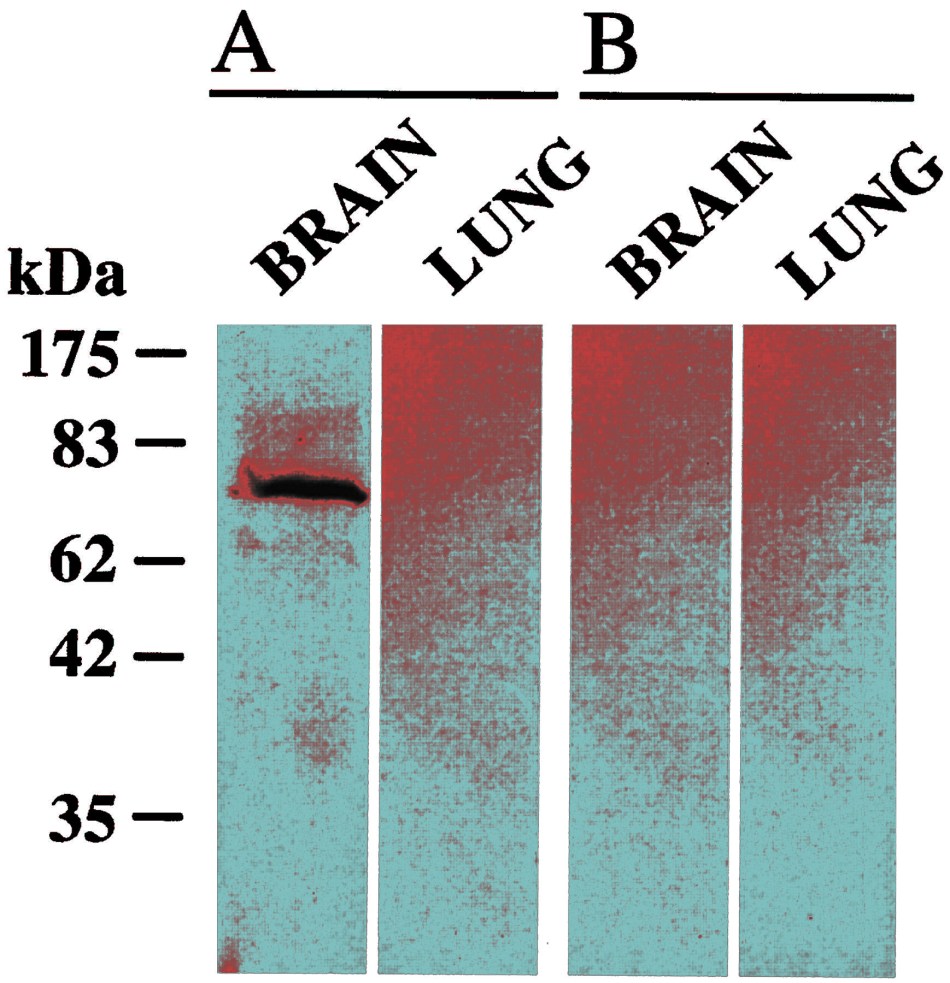
This is the offending Figure 1 again, as flagged in October 2016 on PubPeer:
My article appeared on 19 July 2018, and the next day, somebody posted this expert assessment on PubPeer:
“If you study the noise pattern carefully it is apparent that the three images of negative lanes are not the same. They have the same overall appearance but that is more a factor of the method and imaging conditions. It can possibly be three independent images of the same lane but that is impossible to know under these conditions and without raw data. Raw data might be difficult since it is published almost 20 years ago.”
Wrong on all counts, it turned out.
In my follow-up article from August 2018, I mentioned that the first author refused to tell me who generated the offending Figure 1 with its triplicated gel lane. It now turned out, it was she, together with Jean-Pierre Bourgeois, now emeritus research director of Institut Pasteur, who are responsible for the figure.
The corresponding author of the paper, Jean-Pierre Changeux, is 82 years old and was not involved in the investigation (he is still active, in the fall of 2018 he travelled to give a keynote talk in Hong Kong to receive Albert Einstein Award for Science, or to New York to receive the Goldman-Rakic prize). Maybe because of all that travelling the Institut Pasteur decided not to bother their honorary president with boring issues. It was Arroyo-Jiminez who has secured the original 20-year-old data for the investigation. Or so the report says, the investigator Francois Rougeon simply didn’t attach them to his cover letter to KI legal office and to his report. He also eventually replied to Thyberg when the latter asked to see those images, and declared:
“I contact Helena Scarabin to define the best way to settle the Issue.”
Helena Scarabin is KI’s legal counsel, she notified me on 3 August 2018 that KI will not be investigating the case as the work was done in Oslo and Paris. On 20 August 2018, Scarabin wrote to Rougeon asking him if he and his Institut Pasteur “have received notification or information regarding this as well”. 10 workdays later, the institute’s Comité de Déontologie completed its misconduct investigation, an impressive efficiency. However, Scarabin confirmed to Thyberg that Institut Pasteur never shared any images or any other raw data with KI, only the text report and the cover letter. What exactly does Rougeon want to discuss with her then? Also Arroyo-Jiminez did not reply to my request to share the gel image.
In the Rougeon report to KI we read:
“The work published by Arroyo-Jimenez et al results from the cooperation of 8 authors from 4 different laboratories. It is a typical attempt to develop cooperation between laboratories with different and complementary scientific and technological competences. Jean-Pierre Bourgeois, from the Pasteur Institute, went to Oslo for about two weeks to carry out the immunogold labeling observations by electron microscopy following a method originally developed in his laboratory in Oslo and published under fig 9 -11 of Arroyo-Jimenez et al paper under the supervision of Ole Petter Ottersen.”
The report continues (typos theirs):
“The figure published in the printed version of J Neuroscience shows only one band in the left lane with an apparent molecular weight of ca 70kDa. This value is within the range where immune-purified alpha4-subunit protein as has been found by other authors (Whiting et al 1987). Nevertheless, even if the three control lanes in figure 1 appear negative as expected for a specific antibody, PubPeer stated that all three negative lanes are the same.
This comment has led to the allegation of “likely suspected research misconduct”.
This issue has been examined by the Comité de Déontologie from the Pasteur Institute. Using the software “IMAGE J” the analysis confirmed the nearly identity of the three control figures in the published picture.
As a first step in the attempt to understand this unexpected observation, the authors were asked to present, if available, the 20 years old raw data which were at the origin of figure 1. Dr. Maria Arroyo-Jimenez who carried out the experiment in the Pasteur laboratory was able to go back to the original notebooks she had preserved and to communicate a copy of the original photographs of the gels. Despite the rather low quality of the photograph, it is clear that the test lane shows exactly the same band at ca 70kDa which is not present the three control lanes, all this being consistent with the published figure 1 and most of all with the specificity of the antibody.
Yet, the three control lanes show multiple faint bands differing from one lane to the other and corresponding to the background observed in such experiments.
These raw data are thus consistent with the standard specificity test. The issue is then why such background micropatterns do not show up in the published figure?
Dr. Maria Arroyo-Jimenez in her remembering mentions “ we cut the original film to do the final figure” and “I remember we adjusted the grayscale using Photoshop” (practices which were accepted at the time but no today). Thus a possible explanation is that after extensive reduction of the background, for an unknown reason, the three lanes appear the same.
Nevertheless it cannot either exclude that the same lane has been accidentaly used.
In any case, whatever the final explanation of the published image, the evidence for the antibody specificity shown in figure 1 is consistent with the actual raw data.”
It is technically impossible to make different images look same, even after having “adjusted the grayscale using Photoshop“. The Parisian experts can easily pop over to France’s chief investigator, Francis-Andre Wollman at Sorbonne University, even he never thought of that crazy explanation for duplicated bands in papers by Catherine Jessus.
Rougeon also likely knows this “unknown reason” hypothesis makes no sense to sane or to not sufficiently corrupt people, and this is why he added this cited kicker sentence, that the author Arroyo-Jimenez cannot “exclude that the same lane has been accidentally used”.
There you have it. Figure 1 which was generated in the Ottersen lab in Oslo, is obviously manipulated, the same lane was reused three times, accidentally of course. The original data, if it does really exist as such, is not available to nosy outsiders or even insiders like Ottersen’s own university, KI. The rest of the report on why the result was not important anyway or reproduced elsewhere, is irrelevant filler, serving to distract and to bamboozle. Especially this is something which Rougeon and the others can put in their pipes and smoke:
“Moreover, the Arroyo-Jimenez et al 1999 paper has been positively quoted by at
least 19 articles in the past 20 years, without any negative or controversial statement.”
Thyberg himself is an emeritus professor of cell biology at KI, who describes his past work as “focused on electron microscopy (EM)” and declares to have “experience of immuno-EM”, sees the conclusions of Rougeon report as weak. Thyberg commented to me:
“In the CVDC report, it is claimed that the immuno-EM part of the study, including immunoperoxidase labeling (made at the Pasteur Institute) and immunogold labeling (made in Oslo), supports the specificity of the acetylcholine receptor antibody. It is also said that the article has been quoted 19 times in the literature without questioning of the results.
The immuno-EM illustrations are not of good quality with poor structural detail (a frequent problem in immuno-EM) and partly unspecific labeling. For example, in the immunogold labelings, the number of gold particles are few and they are observed not only in postsynaptic densities but also in other locations, including the extracellular space. Perhaps this is the reason why the authors are very cautious in describing the controls made without primary antibody or with primary antibody preadsorbed with the antigen peptide. In these controls they say that “most of the synaptic profiles were unlabeled”.
The paper has been quoted 19 times in the literature over a period of 20 years. This is not a very impressive citation frequency. Of these 19 papers it is only in 2 that occasional immuno-EM illustrations are found. In the first of these papers (Hu et al, 2007), it is only mentioned that they used the same immunogold procedure as Arroyo-Jimenez et al (the paper investigared by the CVDC). No comparison with the results of this paper is made. In the second paper (Brown et al, 2012), an immunoperoxidase method was used to stain mouse brain sections. The results section states that, like in the Arroyo-Jimenez paper, labeling was observed in the endoplasmic reticulum and near the plasma membrane of neuronal cell bodies. However, like in this latter paper, the quality of the illustrations is not convincing.
So summing up, there are basically only two papers in the literature in which immuno-EM labelings of a similar type as in the Arroyo-Jimenez paper have been done. As a whole, they cannot be said to either give strong support or to disprove the results of the latter paper.”
Update 19.03.2019
Thyberg now received an email from Rougeon containing a gel image:

“Dear Dr. Thyberg,
As underlined in the report sent to the Karolinska Institute on September 4, 2018, we do not exclude that the three control lanes of Fig. 1 are the same.
In your mail of March 8, 2019, you asked for the copy of an uncut immunoblot with the positive and the three control lanes in the same picture. Unfortunately, such a document has not been retrieved. Only pictures of two separated original blots are available. You will find them attached.
The two uncut lanes of the left moiety correspond to the part A of the Fig. 1 while the two lanes of the right part correspond to the part B of the Fig. 1.
We agree that these documents do not constitute an irrefutable demonstration of the specificity of the antibodies raised against the alpha4-chain but only a strong presumption. This, however, is unquestionably established firstly by the results presented in Fig. 2 of the same article and secondly by the results reported in Nature (1999) 398, 805-8101, a reference which is quoted in the incriminated paper.
In conclusion, if it is obvious that, whereas the Fig. 1 does not formally establish the specificity of the antibodies, the conclusions reported in this paper cannot be questioned as already pointed out in my report to the Karolinska Institute.
Sincerely, François Rougeon
- Marubio LM, del Mar Arroyo-Jimenez M, Cordero-Erausquin M, Léna C, Le Novère N, de Kerchove d’Exaerde A, Huchet M, Damaj MI, Changeux JP.
Reduced antinociception in mice lacking neuronal nicotinic receptor subunits. Nature, 1999 Apr 29;398(6730):805-10. PubMed PMID: 10235262.”
If one puts Rougeon’s allegedly original data next to published figure, it doesn’t really match. Not even the first lane and the band in it, all four lanes are different between Rougeon’s data and published Figure 1. Yet that allegedly original data served as evidence to dismiss the suspicion of research misconduct and to declare that no data manipulation took place. How often does this happen in whitewashing investigations?
Donate!
If you are interested to support my work, you can leave here a small tip of $5. Or several of small tips, just increase the amount as you like (2x=€10; 5x=€25). Your generous patronage of my journalism will be most appreciated!
€5.00


Ottersen and Macchiarini is the New Karolinska. Sadly the corruption has taken over the home of the Nobel price in medicine. The price in literacy has also collapsed in a corruption scandal.
LikeLike
Pingback: Fd Professor: Uppföljning kring misstänkt oredlighet i forskningen på KI - NewsVoice
Pingback: The Wonderful Adventures of Nils Billestrup with Swedish gels – For Better Science
Pingback: The Redemption of Arturo Casadevall – For Better Science
Pingback: Misconduct findings fell KI Vice-President Dahlman-Wright – For Better Science
Pingback: Karolinska’s haunted leadership – For Better Science
Pingback: Academic throne succession: from Anne Dejean to Oliver Bischof – For Better Science
Pingback: l’Amoura pour la cigarette Changeux tout COVID-19 – For Better Science