
My regular contributor Smut Clyde went potty over nanotechnology, and it might be my fault. The nano-science can sure be entertaining, as much of good fiction literature is, and some speculate nanotechnology might be even useful, or at least self-important enough to call a peer critic a “scientific terrorist”. The lay-blogger Smut Clyde, instead of being a functional member of society and writing articles on how nanotechnology will cure cancer, obsessively patrons PubPeer for anything nano-sized happening. He then writes posts like this here, inspired by expensive art and cheap science fiction smut, for this non-peer reviewed pseudo-journalistic website.
Now Smut Clyde even got help from a social media character in feline disguise, TigerBB, who popularised Smut’s previous nano-installment in Mandarin in the cheater’s native homeland of China, over the micro-blogging platform Weibo. That time it was about Dr Dong Ge Tong, and his Chengdu University of Technology replied that they opened an inquiry and that Dr Tong will be repeating his Photoshop “experiments”.
Because China’s Communist Party announced with their usual bombastic pomp a crackdown on research fraud, Smut Clyde, together with TigerBB, offered to help. This is why the hero of our story is another Chinese Photoshop-enthusiast, Rijun Gui, a “specially recruited professor” at the School of Chemistry and Chemical Engineering of Qingdao University in the Shandong Province, China. There is also a female lead, Gui’s wife and colleague Hui Jin. The two nano-explorers have a co-authoring boss, Zonghua Wang, and second-degree-affiliates like Lianjiang Tan, who like Gui was once postdoc the lab of Ajun Wan at Shanghai Jiaotong University. Almost 30 of their papers, mostly published in for some reason respectable Elsevier journals, are being discussed on and by PubPeer, one was already retracted by the Royal Society of Chemistry.
I personally want to help world’s most loved publisher Elsevier here, even if they declared me a “toxic individual”, on another nanotechnology and material science-related occasion. And now Elsevier even blocked me on Twitter and set their so-called “Director of Scholarly communication” upon me.
![Do5S_RiX0AAcgLh[1]](https://forbetterscience.com/wp-content/uploads/2018/10/do5s_rix0aacglh1.jpg?w=278&h=230)

But back to our Chinese cheaters, and their Elsevier achievements. TigerBB assembled a handy list of the characters involved and of their problematic papers, and already introduced Chinese audience to this Qingdao team of Photoshoppers over Weibo:


Now, Smut Clyde tries same, but in English.
Treachery of Images, by Smut Clyde
Three years ago, RetractionWatch reported on a leading candidate for the hotly-contested title of Year’s Most Opaque Retraction.
Of the original nine authors, six denied any role in its publication (including the Corresponding Author, who had presumably submitted the manuscript to Chemical Communications and handled the revisions without noticing): “they did not participate in the experimental research that was reported in this article“.
A seventh, Xifeng Liu, was involved only to the extent of improving the English, which he did not think should be dignified with co-authorship. Dr Lin is currently affiliated with the Mayo Clinic. The other non-authors’ institutional affiliations were merged together in an undifferentiated welter, covered by a superfix ‘a’.

That left Rijun Gui and Hui Jin, who had lost confidence in the paper: they complained that results in the Online Supplement had been copied from elsewhere, and that they were unable to reproduce the experimental results or follow the calculations in the main body. It remained unclear who (if anyone) was responsible for inserting the plagiarism, false data, or faulty equations. Nor was it clear who forged the signatures of the seven non-authors. At any rate, they bore no grudge for the theft of their names, and they continued to contribute to later Gui / Jin publications (unless those later appearances were also involuntary).
The paper subsequently attracted criticism in the form of a PubPeer thread, because the stated reasons for retraction were far from exhaustive. Close examination of Figure 1(a) (“TEM … of the as-prepared NS-GQD”) reveals the putative “graphene quantum dots” to be Photoshop clones.
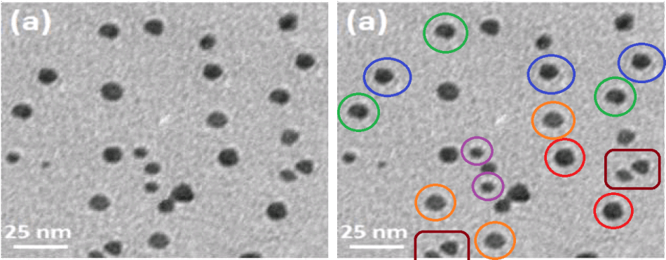
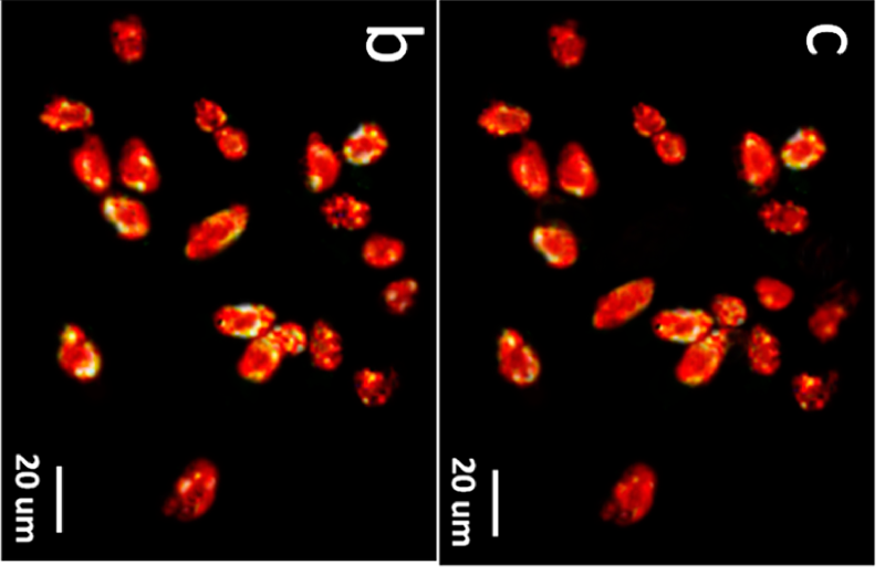
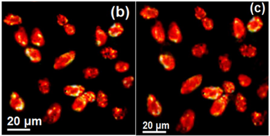
The lipstick kisses of Supplementary Figure S7(a) purport to show a tissue culture of HeLa cells which have absorbed the GQDs, causing them to fluoresce on exposure to 800nm photons… so it causes concern that the same cells also appeared in Jin, Gui, Sun & Wang (2016) as Figure 5(d), where they were identified as “HeLa cells after incubation with Ag2Te@SiO2-NH2 (0.1 mg mL-1). Images were acquired upon excitation at 808 nm”. Not just different nanoparticles, but an altered configuration.
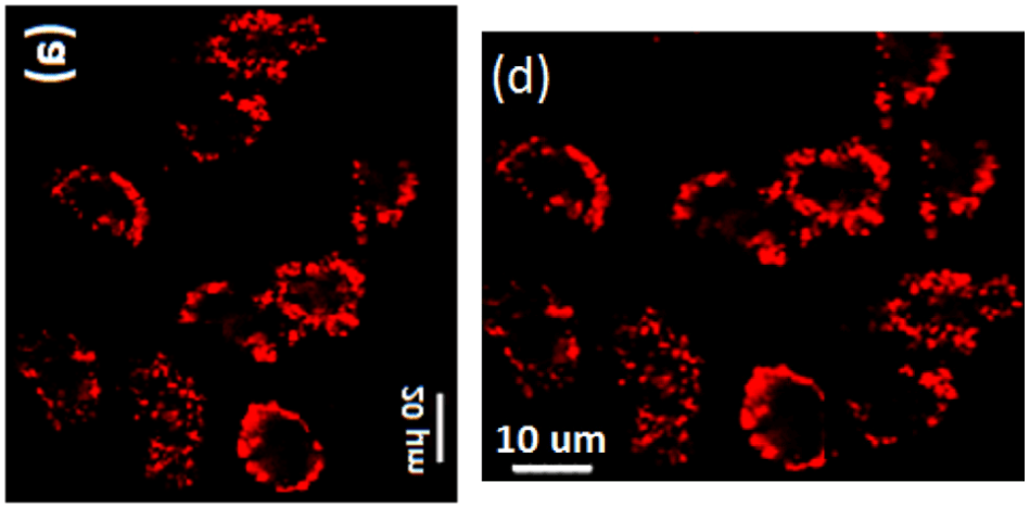
Which is to say, one or both Figures is fake, and might just show some variety of sea anemone. And from there the vista expands to an entire oeuvre of papers, covered by interlinked threads at PubPeer.
Readers will be thinking “So what, another charivari of chicanery from the Nanotech literature”. But there are enough novel elements in the case to reward our attention. The papers between them illustrate several of the alleged applications of Nanotechnology, favoured by researchers to loosen the purse-strings of governmental funders. They also feature an unusual phenomenon: duplicate publication of microphotographs or spectrographs (identified as different entities), with no authors in common between the papers in which they featured. To put it another way, either Sheldrake’s Morphogenetic Field was working overtime, or a broad collaborative cartel of authors were drawing upon an image bank as communal property.
The authors featured in this oeuvre of PubPeer threads (covering 42 papers at the time of writing) fall into three groups:
A. Lianjiang Tan (sometimes Ran Huang; Shuiping Liu; Yu-Mei Shen; others).
B. Ajun Wan; Huili Li; Jie Sun (and a few others).
C. Rijun Gui; Hui Jin (sometimes Xifeng Liu; Min Yang; Zonghua Wang; others).
There are authorship lists combining only Group-A names, or only Group-C names, but names from Group A and Group C never overlap. Group B are the ‘bridge’, not appearing in the oeuvre on their own, but collaborating sometimes with Lianjiang Tan (and others from Group A), and sometimes with Rijun Gui and Hui Jin (and others from Group C) – never both at once. Perhaps the three-fold division represents departments within Shanghai Jiao Tong University.
I have been advised that having gained his PhD in 2012 (with Xueqin An as advisor), Rijun Gui did a post-doc under Ajun Wan (at the School of Chemistry and Chemical Engineering, Shanghai Jiao Tong University). Ajun Wan’s “bridge” position is explained by his earlier supervision of Lianjiang Tan as a previous post-doc. It further simplifies the dramatis personae to know that Hui Jin and Rijun Gui are married, while Zonghua Wang (also from Group C) heads the laboratory.
“TEM time is expensive?”
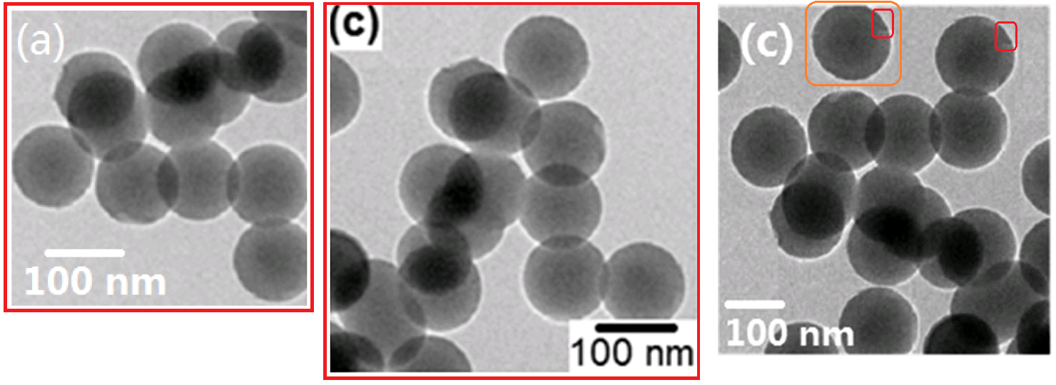
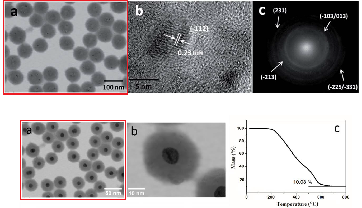
Gui and An (2013) wrote of “Layer-by-layer aqueous synthesis, characterization and fluorescence properties of type-II CdTe/CdS core/shell quantum dots with near-infrared emission“. The coauthor here is Xueqin An, Gui’s PhD supervisor. Despite appearances, the panels of their Figure 3 are not maps of a mountainous archipelago, from a five-volume series of fantasy novels.
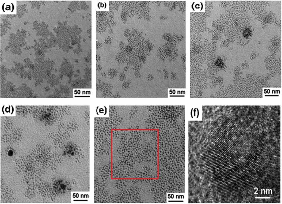
In fact they applied transmission electron microscopy to swarms of the freshly-synthesised quantum-dot cores, and their progressive expansion in a sequence of layers: “Wide-field TEM images of CdTe core QDs (a), the corresponding CdTe/CdS core/shell QDs with 1 (b), 2 (c) and 4 (d) MLs of CdS shell, TEM (e) and HRTEM (f) images of CdTe/CdS/ZnS core/shell/shell QDs with 4 MLs of CdS shell and 2 MLs of ZnS shell.”
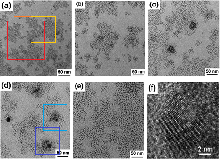
Largely-overlapping enlargements appeared as “PEI-stabilised Carbon QDs” in Figure 2(a) of Gui et al (2014a), and as dopamine-conjugated CdTe quantum dots in Figure S1(a) of Gui et al. (2014b). To explain how details of these panels were blown up, rotated and relabelled as different materials, across a long series of papers, PubPeer contributor “Epichloe Festucae” could only surmise that “TEM time is expensive?“.
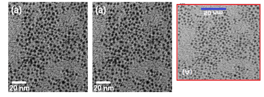
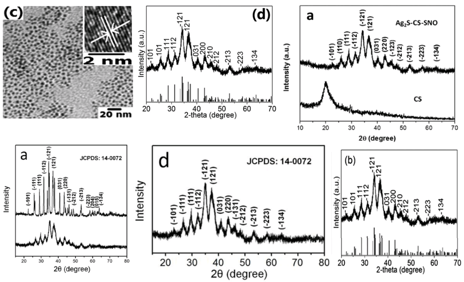
And at right, after a four-year delay and a vertical flip, as “CDs prepared from hydrothermal carbonization treatment of fresh black fungus juice“, as Figure 3(a) of Jin et al. (2018). By then the journal peer-reviewers were expecting the strings of meaningless buzzwords to be longer, so these carbon dots “facilely self-assemble” with magnetic nanoparticles and aptamers to form tetradotoxin-detecting nanocomposites.
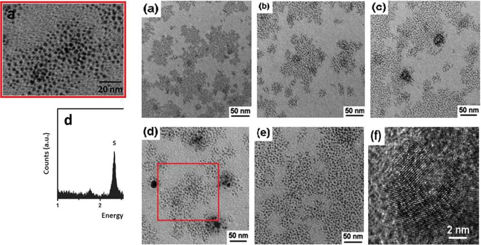
In Figure 1(c) of Gui et al. (2014c), the panels became “Ag2S QDs”.
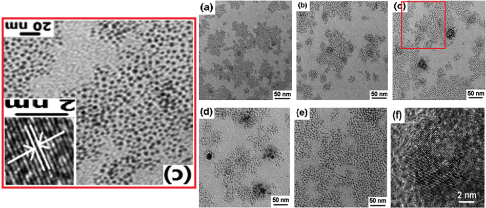
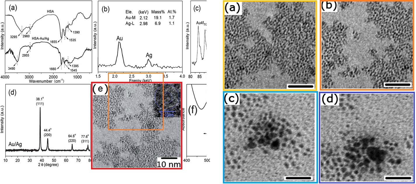
Space does not permit demonstrations of all ten papers repurposing the 2013 Wall of Dots. We must be content with Gui & Jin (2013), where the particles were “human serum albumin-stabilized fluorescent Au/Ag core/shell nanocrystals for highly sensitive and selective sensing of copper(II)“, illustrated with five enlargements from the original;
And as Ag2S QDs again, as Figure 2(a) of Tan, Wan & Li (2014): one of two “Group-A” papers, accessing the original image despite an absence of authorship overlap with the other eight papers in this series. The second being Tan et al (2014), where the QDs became Ag2Se and were capped with multidentate polymer.
These were not the only figures weaving these papers into a single body of work. Other cross-connections caught the eyes of commenters.
These overlapping circles are either “blank liposomes”, Supplementary Figure 3(a); or “mesoporous silica nanoparticles (MSN)”, Figure 1(c); or “CMC–FA–RBS hybrid nanospheres”, Figure 2(c). But who knows? They could be something else entirely.
As is always the case when reporting on Nanomalfeasance, I am cherry-picking the Pubpeer threads, picking out only a fraction of the coincidences, in accordance with obscure aesthetic criteria. Other people’s tastes might differ (for instance, the PubPeer twitter account), so readers are urged to explore the original sources.
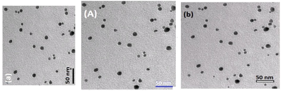
We can widen the investigation by going back to Tan, Wan & Li (2014). The gist of that paper was that when HepG2 cancer cells are fed a diet of silver nitrate and sodium sulfide, they synthesise Ag2S ‘quantum dots’ within their cytoplasm (through the mediation of reduced glutathione). The photofluorescence of these QDs betrays their presence when they are extracted from the hepatoma cells and injected into mice. Here, at left, I have flipped Figure 4 horizontally [left]…
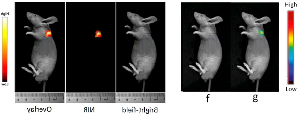
… to highlight its resemblance to the mouse [at right] from Figure 7 of Tan, Wan & Li (2013). Though that mouse was glowing in the dark (irradiated by 488 nm laser light) because it had been injected with CuFL and Ag2S-GSH-SNO nanoparticles. It is a highly-trained Stunt Mouse with experience in this act; readers should not try it at home.
The HepG2 cells themselves are depicted in Figure 1, in typical scenes from a tissue culture, increasingly luminous at “(d) 12, (e) 16, and (f) 20 h after uptake of silver nitrate and sodium sulfide” as they migrate across the culture plate.
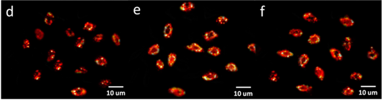
They could be goldfish, painted by Paul Klee. They could also be the HeLa cells featured in Figure 4(d) of Jin et al. (2016), glowing because they had absorbed Ag2Te@SiO2-FA from their culture medium (though sometimes rotated through 90º). We met Jin et al. (2016) earlier, where the glowing HeLa cells formed a rockpool gallery of sea-anemones.
I love Klee goldfish so one more sample before moving on! You will recall that Tan, Wan & Li (2013) were “Conjugating S-nitrosothiols with glutathiose stabilized silver sulfide quantum dots for controlled nitric oxide release and near-infrared fluorescence imaging“. These conjugated nanocomposites not only made mice glow; they had the same effect on another line of cancer cells. Thus, Figure 6, “(b) NIR image acquired immediately upon 808 nm irradiation, and (c) NIR image acquired after 808 nm irradiation for 2 h of L929 cells incubated with 0.1 mg/mL Ag2S-GSH-SNO nanoparticles for 3 h. … ” Here rotated through 90º…
…to facilitate the comparison with Supplementary Figure S7 of Gui et al. (2014d), where the Ag2S quantum dots were “highly stable and biocompatible” (I have flipped them horizontally due to my Gerstmann’s Syndrome). This is another of those “no authors in common” situations.

Figure 5 of Wang et al. (2015) also cries out for comparison. By 2015, the fluorescing quantum dots were “Ag2Te … with compact surface coatings of multivalent polymers“.
Despite his youth, Rijun Gui is a prolific worker, with Group C collecting 28 Pubpeer inquiry threads. Group A is less prominent (only 14 queries for Lianjiang Tan) but we should not neglect it. Tan’s work has taken a slightly different direction, pursuing nanoparticles as possible drug-delivery vehicles – especially as nanopackaging for targeted chemotherapy – more than as cytotoxic cures for cancer in their own right. In general his use of retouching is more sophisticated.
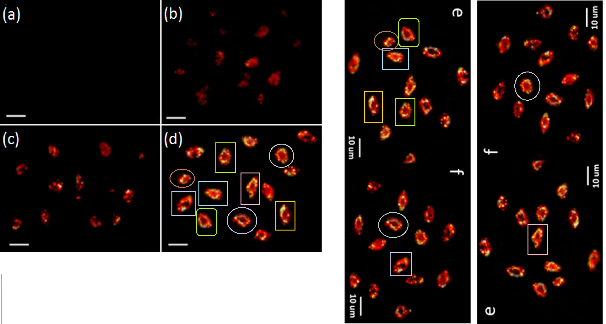
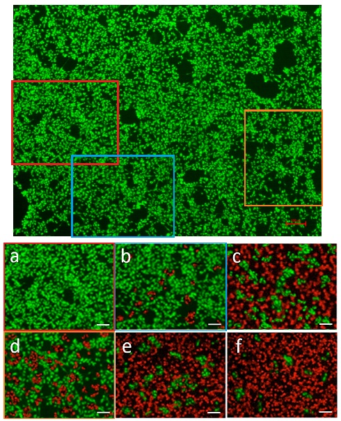
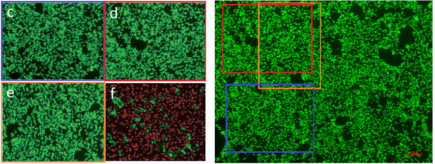
Notably, in the “wall of green” below, the upper half is Fig. 4 from Yang et al. (2015): “Representative fluorescence image of NIH 3T3 cells after incubation with PEG5000–PLA5000 micelles of 1.0 mg mL-1 for 48 h. (The cells were stained by AO and EB.)”
The lower half is Figure 7 from Tan et al. (2017): “Fluorescence images of CT-26 cells with live-dead staining after treatments with different formulations: (a) PBS; (b) DOX; (c) UCNPs(DOX)@CS; (d) UCNPs@CS-RBS plus 30 min; (e) UCNPs(DOX)@CS-RBS plus 5 min; (f) UCNPs(DOX)@CS-RBS plus 30 min. The living cells were labelled by calcein AM (green emission), and the dead cells were labelled by propidium iodide (red emission).” The coloured rectangles indicate matching arrangements of NIH3T3 and CT-26 cells… the latter modified by adroit replacement of green dots with red ones. The other three CT-26 panels may have NIH3T3 counterparts as well, but too modified to be recognisable.
A similar phenomenon occurred with Figure 6 of Tan et al. (2015), at left below: “Fluorescence images of HeLa cells before and after irradiation by 980 nm laser with the power density of 0.5Wcm-2 over a period of 5 min in the absence (c and d) and presence (e and f) of the CuS mesostructures with the concentration of 0.3 g L-1).” That time, four panels could be recognised within the larger wall at right.
These images invite comparison. Left is Figure 1(a) from Tan et al. (2012): “TEM micrograph of CdTe QD–CMCS nanocomposite NO donors [horizontally flipped]. Right is Figure 1(c) from Tan et al. (2014b): “TEM image of Mn2+–ZnS@CS-RBS.”

Then there was a pair of papers in which Tan demonstrated how to turn a electron-microscopy scene into a congeries of googly eyes (or frog-spawn, it may be), using the photoshop equivalent of a crayon. The papers were Tan et al. (2014c) and Tan et al. (2017b). The Before and Frog-Spawned images are the respective Figures 2: “(a) TEM micrograph and (b) high-resolution TEM micrograph of Ag2S QDs@CS-RBS nanospheres. (c) SAED pattern of Ag2S QDs@CS-RBS nanospheres”; and “(a) TEM image and (b) high-resolution TEM image of Ag2S(DOX)@CS nanospheres. (c) TG curve of Ag2S(DOX)@CS nanospheres.”
I was going to finish there, but there remain these X-Ray Diffraction patterns, displaying a mastery of laboratory technique capable of replicating the finest details of noise… especially as the source materials are variously identified as Ag2S-CS-SNO nanospheres, Ag2S QDs prepared in different ways, or Ag2S aggregates before calcination. They come from disjoint groups of authors: Gui et al. (2014c), Tan et al. (2013), Tan et al. (2015), Tan et al. (2017), and Gui et al. (2014d).
Connoisseurs of the genre may also enjoy these wobbly, overhanging zeta functions that appear to have been left out in the sun too long, so they started to melt (Figure 1(f) of Xu et al. (2017)), at left; and this NMR spectrum, in which the baseline is composed of repeated sections, and a repeated / mirror-imaged section (Figure S2 of Tan et al. (2015)).

Early in 2018, it was asserted through social media that Rijun Gui regrets his actions and intends to resign his University position.

This has not yet been confirmed, and he is hardly alone in bringing shame upon the University. It would also leave the papers in circulation… the journals involved have shown no enthusiasm for retracting them. Until further developments, here are some recent examples of replicated and rotated images:
The nanodots are either Fe3O4 NPs (in Jin et al. (2018)); CuS NCs (in Xu et al. (2017)); or CD2 conjugates (in Gui et al. (2017)).
Donate!
If you are interested to support my work, you can leave here a small tip of $5. Or several of small tips, just increase the amount as you like (2x=€10; 5x=€25). Your generous patronage of my journalism will be most appreciated!
€5.00


Wang et al. (2015) was left out from the main post because it didn’t meet my aesthetic purposes.

Still, it’s worth a look.
IIRC, it was the first paper from Gui’s group to appear in PubPeer.
LikeLike
Let’s keep in the realm of artful Science (maybe that was what Leonid meant anyway):
The adventures of Greggery Peccary (Frank Zappa, written a long time ago, but he was really ahead of his time):
“And what, might you ask, is a TREND MONGER? Well, a TREND MONGER is a person who dreams up a TREND (like ‘The Twist’ -or ‘Flower Power’), and spreads it throughout the land, using all the frightening little skills that Science has made available!”
LikeLike
Not in the field of nano-photoshop but also originated from PR China. This one is a very severe case of clearly intentional self-plagiarism/duplication. One doesn’t need to understand thermodynamics/chemical reaction engineering to grasp how come both pubishers (the beloved Elsevier and RSC) and plagiarism checker could fall to such puny trick.
https://pubs.rsc.org/en/content/articlelanding/2018/ra/c8ra04440f
https://www.sciencedirect.com/science/article/pii/S0360319918315295
Sadly in the field of chemical/energy/mechanical engineering (especially on applied one, not the science/nano-level), publication ethics are clearly not as publicized, exercised, and heralded as that of biomeds.I would say the problem might be just as bad or worse of, because simply no one cares.
LikeLike
Elsevier and other publishers should be boycotted until they show responsibility and stop this fraud mill!
There seem to be a total lack of peer-reviewing and quality control in a number of journals.
One can laugh about the nano fun, but the fake science can also have negative impact on
the society, eg. the disturbed anti-vaccine people use faked or low-quality papers to have an influence.
In France four out of ten are now sceptical against vaccine!
Leonid, have you contacted the Elsevier leadership? I complained to them some weeks ago but didn’t hear back…
LikeLike
My understanding is that Leonid has been boycotted by Elsevier
LikeLike
Certainly! Leonid’s enemies are our enemies!
Regarding predatory publishers: I have started testing new tactics against the daily emails I get with invitations to fake conferences in China: I answered and suggested some topics for my talk. One of them was to improve the human rights and development of democracy in China. It seems to be quite efficient. Suddenly I did not get any more invitations from the Bit conferences.
LikeLike
Many times many PhD students have a special forced PhD course which consists in Adobe Photoshop manipulation of microscopic photos
However sometimes certain specialist journals indeed pick up the manipulations…
LikeLike
Readers are probably bored for the moment with Nanofraud stories from Chinese laboratories. If anyone is not, they could check out Mi-Cong Jin’s group for some Photoshop crimes that are so egregious that even the journal editors have issued a few Expressions of Concern.
https://pubpeer.com/search?q=+Mi-Cong+Jin
Includes googly eyes! (or frog-spawn, as it may be).
LikeLike
The first retraction from this batch:
https://www.sciencedirect.com/science/article/pii/S0039914017308627?via%3Dihub
This article has been retracted at the request of the Editor following concerns raised by a reader.
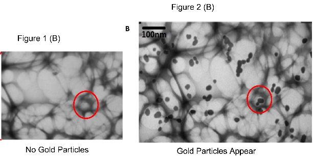
The article uses two electron micrographs which have been used in other publications as well, denoting different samples.
Fig. 1A is identical to Fig. 3E in RSC Adv., 2013,3, 20959-20969 doi: 10.1039/C3RA43120G despite describing different samples.
Fig. 2A is identical to Fig. 1B in Sensors & Actuators B: Chemical, vol 245, pp 386-394 https://doi.org/10.1016/j.snb.2017.01.166 and Fig. 1A in Materials Letters vol 195 pp 131-135 https://doi.org/10.1016/j.matlet.2017.02.119 despite describing different samples.
These problems with the data presented cast doubt on all the data, and accordingly also the conclusions based on that data, in this publication.
The scientific community takes a very strong view on this matter and apologies are offered to readers of the journal that this was not detected during the submission process., http://dx.doi.org/10.1016/j.talanta.2017.08.043.
LikeLike
Probably this was the first. So far, 2 of the 28 of Gui and Jin’s papers with image frauds have been retracted.
https://www.sciencedirect.com/science/article/pii/S0925400517301739?via%3Dihub
LikeLike
Pingback: Carrots and sticks for fraudsters at Royal Society of Chemistry – For Better Science