
The biochemistry research lab of professor Roland Lill at the Philipps University Marburg in Germany is a place where space, time and western blot continuum collapses into an anomalous singularity, where paradoxes abound, but only one fact remains certain: there was never any data manipulation in the lab of this senator of the German Research Council (DFG). Only misunderstandings (here) and solid science, with sometimes unorthodox figure assembly methods (here).
One of those anomalies is the recent correction by Lill and his former PhD student Heike Lange (now tenured CNRS researcher at The Olivier Voinnet Institute for Research Integrity in Plant Sciences (IBMP) in Strasbourg, France, published in the prestigious journal PNAS in March 2018. A western blot was found to contain duplicated, triplicated and mirrored bands, and was replaced with a version of same gel, its irregularities fixed. What exactly the new old figure shows, is not clear. The Ombudsman of Marburg University insists that no digital image versions of the western blot exist, only some almost two decade old thermo-paper printouts which were shared only with PNAS. Neither can those archive documents be scanned or photographed, as it will probably either destroy them, or alter the results they show. A PNAS editor however admitted to me that the correction actually shows a pdf file which the authors Lill and Lange supplied by email, a gel image which again seems to be different from the original thermoprinter records. Yet also this digital pdf file cannot be shared, probably because otherwise the time-space continuum will collapse and our universe, or at least the Marburg lab of DFG Senator Lill might end up teleported back into the year 1999, when the prints were made. And anyway, all two decade old experiments were faithfully reproduced by the Lill lab just now, using exactly same reagents. Those results however are also apparently not for sharing.
This was the offending image, which led to PNAS correction, figure 4B in
A mitochondrial ferredoxin is essential for biogenesis of cellular iron-sulfur proteins. Proceedings of the National Academy of Sciences (2000) 10.1073/pnas.97.3.1050

The evidence appeared on PubPeer in February 2017, yet the first author Lange explained on Twitter:
“We started to think about this correction even before the figure was discussed on Pubpeer, only driven by the motivation to get things right”.
It was actually her third correction with Lill (previous two from October 2017 for Kaut et al JBC 2000 and Lange et al JBC 2004), and this is now the corrected image for Lange et al 2000 as published by PNAS on March 12, 2018:

You might recognise the bands 1-4 as well as band 6 from before, other bands are new arrivals, to replace the mishandled bands 5, 7 and 8. You might also notice that the new image is of rather low resolution and even more background-featureless and contrast-boosted than the previous one. Compare with Figure 2 of the same Lange et al PNAS 2000 paper, which has several perfectly normal western blots, none of them lacks background or shows any irregularities. But the western blots in Figure 3 were for some reason also contrast-boosted, just like Figure 4B, and one does detect some splicing in blots of Figure 3A (since at least some residual background remained).
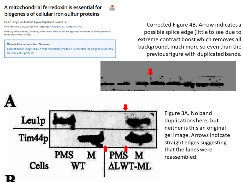
In fact, one even sees traces of possible splicing in the corrected Figure 4B, despite its otherwise featureless background. This was the accompanying Correction notice:
“The authors wish to note the following: “Recently when we reanalyzed Fig. 4B (a Western blot visualizing the yeast protein Leu1), we noticed that there was an error in the assembly of the bands. Because the gel loading for the Western blot did not fit the desired order for presenting the data, the first author cut the thermoprinter paper printout into pieces and reassembled them. Unfortunately, the published version contained an unintentional data duplication (mirrored bands for the 64 h data points), but the overall results were not impacted. We apologize for the mistake.” The corrected Fig. 4 and its legend appear below”.
Scissors, paper, glue
It is a bit puzzling why, if Lange saw the need to rearrange lanes to get them into a certain order, exactly same sample order is shown in the corrected figure. In any case, one still wonders how cutting and reassembling a gel printout can lead to mirroring of bands. Photographing an X-ray film upside down would flip the bands vertically (which we do not see), but not horizontally (which we do). Unless one took two sets of images, with the transparent X-Ray film flipped and photographed from the front and back, printed both, in several copies, and then attacked those with scissors and glue. But why on Earth would anyone do that?
According to Lange writing on PubPeer, only one or two copies of that printout must have existed in the Lill lab in 2017:
“The thermoprints were then cut and glued on the paper. I generated a sufficient number these cut-and glued print layouts for each figure: one for the journal, two or three for review, one or two for us to be kept”
Which means: one of those was sent to PNAS last year, one last version remains with Lill. Yet neither was photographed or scanned, as declared. This was the statement I received from Daniel Salsbury, Deputy Executive Editor at PNAS:
“The author contacted PNAS by email at the end of 2017. He included an explanation for the correction and a PDF for the corrected image. We also received a hardcopy version of the same information later in January. The highest resolution image is available online in the HTML or PDF of the correction: http://www.pnas.org/content/early/2018/03/06/1803134115.”
After the correction, Lange explained on PubPeer what happened, indicating that indeed a digital version of a correction file, “a scan”, exists:
“2. How the error likely occurred
In this particular case, the print template did not fit to the size the bands have on the X-Ray. In addition, the X-ray showed additional control bands that were not mentioned in the text and had no place in the template. Hence, I cut the bands that should be shown in the figure individually and glued them in the necessary order to the template. Once the bands are arranged to a stripe, I photographed them again for size adjustment, and to avoid gluing 8 single bands 5 times. I assume that this explains why the splice sites are not obvious, but you can probably detect them with forensic software. Obviously and undenied, the procedure is not idiot safe and in fact a recipe to make errors. It happened that I put the X-ray upside down in the camera box and cut bands without noticing it. This results in mirrored bands. It happened that I picked twice the same band, which results in duplicated bands. It happened that I printed multiple copies of a wrongly assembled stripe, throw them away {or not:-( } and started from scratch. The published figure received one of these wrongly assembled stripes without that I noticed the mistake.
- What is seen in the correction
The original X-rays were thrown away when I moved into a new flat with little storage place about 4 years ago. At that time I have kept them for 10 years without ever going back to them, and I honestly assumed that I will never need them again. Therefore, I can not provide the original data. What comes as close as possible are two of the old paper printlayouts from 1999 that we found in the offices archive. Both are identical and do not show the duplicated bands of the published figure. We send them per real mail, as paper, to the journal. In addition, we had the experiment repeated by a person who was not involved at the time, and these new data were also send. The journal did not take the decision easy, but they finally accepted this for a correction. The corrected figure is a scan of the paper print layout from 1999 as it was send to the journal. As it is no digital remake, it shows the thermoprint stripe assembled from cut bands without that the splice sites were signaled as one would do today”.

Dark matter
The last part contains several peculiar bits of information. First of all, the original research data in Lill’s laboratory sometimes behaves like the dark matter. It seems to exist, or maybe not, yet its quantum presence can be detected by the gravity it exerts on the observer. It is not at all normal that a PhD student takes all original data (X-Ray films and who knows what else) with her after graduating, transfers it to Strasbourg, France, eventually dumps it into garbage, while leaving her boss back in Germany with only some thermoprinter copies. In fact, this kind of data removal from the lab is actually forbidden by rules of good scientific practice, not just nowadays, but always. To be on the safe side, in December 2017 the Marburg University issued a guideline that original research data must be stored for 10 years.
However, in other cases Lill’s original data remained safely stored at the university, and Lill himself recovered it and had it posted it on PubPeer (here and here, the latter paper even co-authored by Lange). On the other hand, also the original data for Lange et al JBC 2004 paper was according to the December 2017 correction notice “no longer available“, not even thermoprinter backups. Lange probably took all those also and trashed everything when moving flat. Luckily the authors were able to recover ~15 year old yeast strains and even antibodies and reproduce the experiments faithfully, in order to issue a correction for a flipped and duplicated western blot image. Most other scientists would have difficulties reproducing such old data (aside of Olivier Voinnet, obviously), even if they recovered the ancient reagents unspoiled, but that was no trouble at all for Lill. Time apparently does not move linearly in his lab. Also, what Lill and his former lab members understand with “same” or “identical” is also subject to the cosmological uncertainty principle.
For the not yet corrected Kaut et al JBC 2000 namely, Lill provided a replacement a problematic western blot with a clearly different one, maybe a repeat experiment. That in itself would not be so eyebrow-raising if Lill did not use the new picture as scientific proof that the old one contained no irregularities, because “All bands match the accepted version of the figure“. Which is clearly not true: they don’t match at all.

One is rightly confused what Lange’s scissoring rationale of “print template did not fit to the size the bands have on the X-Ray” might have meant. Do gel bands on an X-ray film expand, shrink or move when photographed? In Marburg, maybe. Were those allegedly specific gel bands of different molecular weight, and needed to be aligned as if it was same? But what kind of science would that be?
And once again, one wonders if the picture which Lange published as correction is one of the two remaining original unadulterated “old paper printlayouts from 1999“, why did she have to rearrange those “because the gel loading for the Western blot did not fit the desired order“, if the order remained exactly the same with the original, fabricated and corrected versions? The only logical explanation is that the corrected figure which is a digital “scan of the paper print layout from 1999” is either also reassembled (as indicated by image above) or shows some other experimental samples and not those described by both old and new figure legends.
Under investigation
If the latter, the correction is not honest and must be retracted and replaced. If the former, authors should indicate the lane splicing they performed now. In any case, under the circumstances it should not be impolite to ask the authors to share that just months old scan or pdf which they sent to PNAS, given that the published image is stark contrast-boosted and of low resolution, precluding almost all image forensics. Yet neither authors nor PNAS are willing to oblige. This is why I asked the Ombudsman of the Marburg University, Prof. Dr.med. Dr.phil Dr.h.c. Helmut Remschmidt, 79 year old emeritus professor and a highly respected figure of authority in paediatric psychiatry.
I originally communicated the evidence on my site to Remschmidt as well as to his Ombudsman colleague, the radiologist Rita Engenhart-Cabillic, and their university on July 19th 2017. After a reminder, I eventually received an email reply on August 15th, where Remschmidt announced to me that “the investigation is in full swing“. When approached about the original image used for the correction in PNAS in March 2018, Remschmidt explained in his email from April 6th:
“In fact, it is an original copy of the manuscript submitted at that time, of which Prof. Lill found five identical copies in his documents for this publication during his investigation of this case. He personally presented to me these original templates (with a thermal printout of the blot) long time ago and I ascertained that these prints came from the year 2000. Such original templates were then used to submit the figures. Obviously there was an error in the submission to PNAS in the submitted picture by the first author Dr. Lange, which was not recognized because of the similarity between the correct and the wrong figure. Electronic original data or films of the 18-year-old experiments do not exist any longer, since they were destroyed in 2015. This situation explains the perhaps ambiguous statement made by Dr. Lange, that the data does not exist anymore. However, this gives no reason to doubt the correctness of the above-mentioned original template. This is even more so since Prof. Lill’s group has recently fully reproduced the criticized data. Me and the journal PNAS were also presented with this new, reproduced data. I am sure that PNAS has carefully reviewed the case before accepting the correction”.
So where Lange said there were merely two last copies of the thermoprint picture, Remschmidt saw five. Neither did the Deputy Executive Editor at PNAS mention anything about receiving any new experimental data, maybe he forgot. Most importantly: Remschmidt is also in conflict with PNAS‘ (and even Lange’s) statement about a pdf of a correction figure sent by email, when insisting that there are not digital files, just original printouts.
I was also wondering who is actually in charge of that Lill investigation, aside of the 79 year old emeritus and psychiatry expert Remschmidt, because his name was the only one featuring so far in the context. It is not really the case that all German universities take evidence of research integrity concerns seriously. In a minor case with University of Osnabrück, I was underhand threatened by the Ombudsman, while Hannover Medical School (MHH) for example bowed to order of its president NOT to investigate the horrendous ethics breach and dishonest claims in the MD doctorate thesis of their own certified trachea transplanter Philipp Jungebluth.
Eventually, the Marburg Ombudsman explained to me the procedure at the Philipps University Marburg:
“1) Philipps University (PU) has a “Standing Commission to Investigate Misconceptions of Scientific Misconduct”, which meets periodically to discuss all the more complex issues. If necessary, it also seeks expert opinions and conducts hearings. The commission has 3 professors and 2 scientific assistants. Members of the legal department also regularly attend. The Ombudsman is a non-voting member of the Commission, which also provides him with support.
2) The PU has adopted “principles and procedural rules for dealing with scientific misconduct”, which are followed, as well as “procedural principles of Ombusperson”.
3) in more complex cases, including the one you complained about, ad hoc committees consisting of highly qualified university professors are formed, with appropriate knowledge and experience in the field under investigation. The report drawn up by the respective commissions will be forwarded to the DFG for comments after appropriate discussions. All of this has happened in the case of Lill. However, the comments of the DFG have not yet been received.
4) In order to inform the students at the beginning of their studies about ethical principles and good scientific practice, the “Marburg Research Academy” (MARA) regularly invites the Ombudsman, who deals with the relevant topics in German and English.
5) The ombudsman draws up an annual report on his activities and presents them to the academic senate.
6) Ombudspersons at PUM have been retired professors for the past decade, all of whom have held important positions for years at the DFG and other research funding organizations. The deputies were each experienced university professors who had not yet reached the age limit”.
The German Research Council DFG is apparently investigating their own Senator Roland Lill, while his past lab members Lange and Balk provide on PubPeer the most outlandish explanations why they had to utterly unnecessarily manipulate data, in Photoshop or with paper, scissors and glue. The scientific results remained always unaffected, also because such practice was perfectly justified scientifically. At least the following case was apparently solved even before the Marburg and DFG investigations began, flagged on PubPeer in March 2017:
The following comment appeared on PubPeer on April 6th 2017. What will Remschmidt and his Marburg colleagues make of this, one wonders:
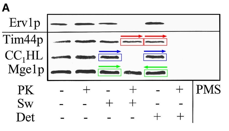
“We have inspected the claim that several bands in Fig. 4 of our EMBO Rep paper from 2001 “are duplicates (red, blue) or flipped duplicates (green)”. Especially in larger magnification, it easily becomes clear that the criticized couples are NOT identical. We further note that the result of this figure (i.e. Erv1 is a protein of the mitochondrial intermembrane space) has been verified by other labs many times since our publication.
Sincerely,
Heike Lange and Roland Lill”

Donate!
If you are interested to support my work, you can leave here a small tip of $5. Or several of small tips, just increase the amount as you like (2x=€10; 5x=€25). Your generous patronage of my journalism will be most appreciated!
€5.00


Pingback: Boletim de Notícias: Avança no Senado PL contra alerta transgênico em alimentos | Direto da Ciência
This is the reflexion of a period of science when rewarded scientists think about and sign science works like pieces of art reflectig their ideas about what the data they should obtain reflecting personal ideas and concepts and not the interpretation of the true, real data
LikeLike
Pingback: Flawed cytometry of Rector Giorgio Zauli – For Better Science
Pingback: DFG and Marburg drop misconduct investigation of Roland Lill papers – For Better Science
Pingback: Spanish elites rally in support of data manipulation – For Better Science
Pingback: Leopoldina and Tiwari’s scamferences, or what’s the point of Academies – For Better Science