
What is it with ophthalmology, are they all blind to see their own fake figures? Sholto David uncovered a totally bizarre case from Germany, from the lab of Professor Stephanie Joachim of Ruhr University Bochum.

Joachim’s boss, the director of eye clinic, is on almost all of these ridiculously manipulated papers. Several feature another important Bochum professor and his son. The investigation of these papers will be fun, because the Dean of the Bochum’s medical school is another artist – Andrea Tannapfel, whose own bad papers on cancer research Sholto exposed just a few months ago.
Joachim once wrote a paper titled “The path to professorship“. Sholto now provides the missing illustrations for it.
Luck in Sight
by Sholto David
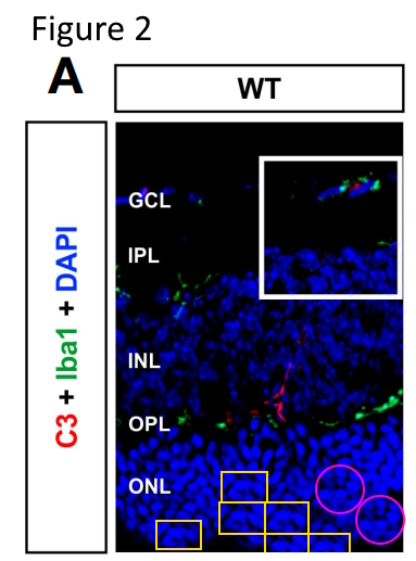
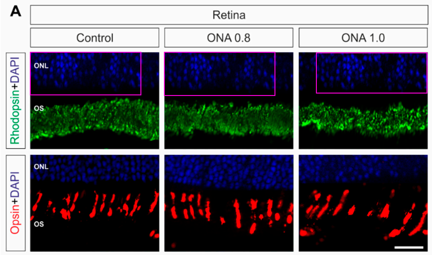
Recently I have been looking at papers in Scientific Reports again. Here’s one that was published in October 2025 (just a couple of weeks prior to the time of writing). There are four pairs of overlapping images that should show different samples.
Sabrina Reinehr, Julia P. Zehge, Katharina Klöster, Maike Mueller, Hasan H. Hendek, Michael Sendtner, H. Burkhard Dick, Ralf Gold, Stephanie C. Joachim, Simon Faissner Retinal degeneration driven by brain-derived neurotrophic factor deficiency in microglia and T-lymphocytes Scientific Reports (2025) doi: 10.1038/s41598-025-21423-6

I shouldn’t bother grumbling about Scientific Reports again, suffice to say it is Springer Nature‘s implementation of the Heliyon cash grab and should suffer the same fate. Speaking of cash, the paper reports no specific funding for the experimental work, only mentioning how they paid the APC (Projekt DEAL).
Most of the authors on this paper are affiliated with the Ruhr-University Bochum (RUB). The last author is Simon Faissner who studied and did his PhD at RUB, then returned in 2017 as a group leader before becoming a full professor of neuroimmunology six years later. His research speciality is multiple sclerosis, exactly what a certain (much older) RUB professor named Andreas Faissner studies. When contacted, nobody denied that Simon is indeed Andreas’s son. We shouldn’t make fun of Italian universities then – German ones are not much better. Faissner Senior has his own PubPeer record, some of it was discussed in March 2024 Shorts.

Tannapfel, a German success story
The papers are old, raw data unavailable, one can see small differences, some students probably did it, and anyway, conclusions are unaffected.
After searching through a few connected papers I decided to look more closely at Stephanie C. Joachim who is the second to last author of the paper. The first author Sabrina Reinehr is Joachim’s former PhD student and still a member of her lab. Joachim herself has the rather lofty title of “Head of the Experimental Eye Research Institute”, although, as is custom in German university medicine, the entire institute seems consist of a medium sized laboratory group. I was surprised to discover many of her papers include carefully and persistently manipulated images published over the last decade. None of these studies have previously been criticized for image problems. I have three main concerns about the images in Stephanie’s papers:
- Duplicate or overlapping images which should show different samples (as shown above).
- Images where the clone tool has been used to duplicate parts of an image, mostly patches of cells, or individual cells.
- Images where the paintbrush tool has been used to obscure parts of the image by “painting” over areas.
I won’t spend too much time on concern (1) which has already been adequately demonstrated and is usually easy to identify. You can see an example of concern (2) below, from the same Scientific Reports paper mentioned above.

This is rather a subtle example and is quite difficult to see. Certain Nobel Prize winners might come up with a whole range of excuses, but I would like to see an escape from the issues in this 2024 paper published by Frontiers in Immunology, here’s a close-up of Figure 2A showing cloned areas in the yellow and pink shapes.
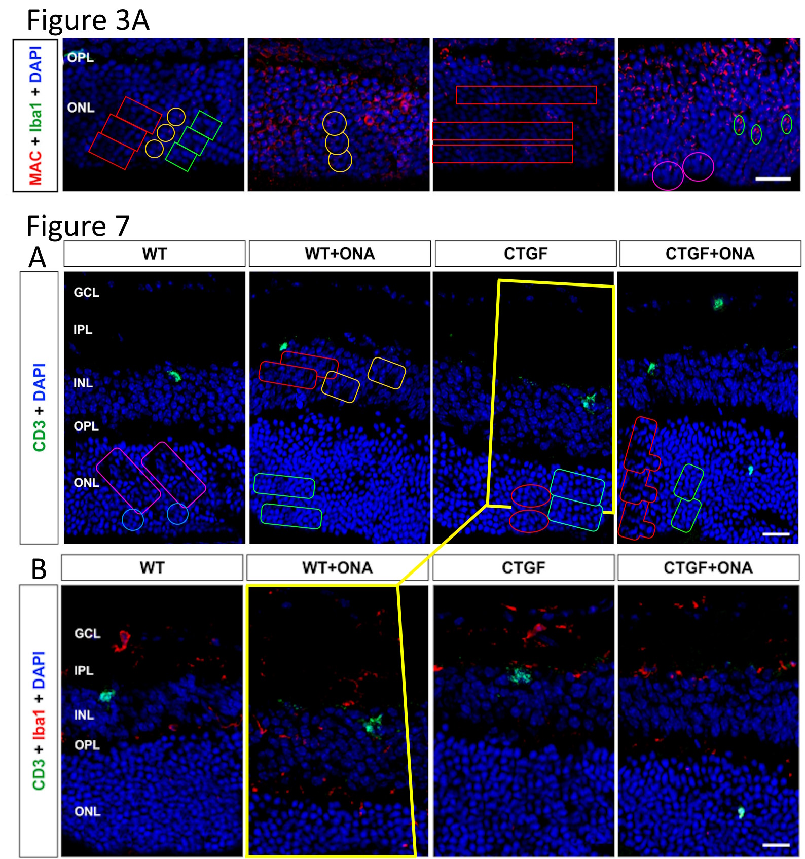
Sabrina Reinehr, Julien Wulf, Janine Theile, Kim K. Schulte, Marcus Peters, Rudolf Fuchshofer, H. Burkhard Dick, Stephanie C. Joachim In a novel autoimmune and high-pressure glaucoma model a complex immune response is induced Frontiers in Immunology (2024) doi: 10.3389/fimmu.2024.1296178
Focus on the yellow rectangles and pink circles. I think the shapes inside the yellow rectangles look like little croissants. The white rectangle is part of the original figure.
Here are some further issues in Figure 3A and Figure 7A from the same paper. I’m sure there are many more cloned areas but after a while drawing shapes becomes tedious. The yellow shapes highlight a large overlapping area between two different samples; I think 7B is likely the original, and all of the extra area outside of the yellow shape in 7A was probably generated with the clone tool.
Unlike in Scientific Reports, this mouse study in Frontiers did use some research funds, from the German Research Council (DFG) and the German Ophthalmological Society (DOG), via a “Glaucoma Research Prize” to Reinehr, who now also serves as social media representative and spokesperson for their program “Young DOG” (not a translation!). Joachim is not a Young DOG though: she chairs the Big DOG board for glaucoma.
Another study sponsor was the local Bochum charity called “Glück im Blick” (German for “Luck in Sight”), which sponsors eye research. Specifically, the board’s own research: the president is RUB professor and director of university’s ophthalmology clinic Burkhard Dick, the treasurer is Stephanie Joachim, and Sabine Reinehr is responsible for social media again. As you can see on PubPeer, Dick stuck his Burkhard into almost every problematic paper by Joachim, and he probably regrets it now.

My Big Fat Greek Ophthalmology
From fake cancer research to fake ophthalmology – just follow Mitsi and Vassiliki and you’ll meet Dementios and other bad eye doctors, including a horrible German we hoped to never see again.
Here’s another paper with cloned areas published in a different Frontiers journal, also in 2024. The mouse research here was funded by the PRO RETINA – Foundation for Prevention of Blindness and the 2023 Dr. Gaide AMD award from the German Retinological Society to Joachim.
Natalie Wagner, Teresa Tsai, Sabrina Reinehr, Janine Theile, H Burkhard Dick, Stephanie C Joachim Retinal debris triggers cytotoxic damage in cocultivated primary porcine RPE cells Frontiers in Neuroscience (2024) doi: 10.3389/fnins.2024.1401571
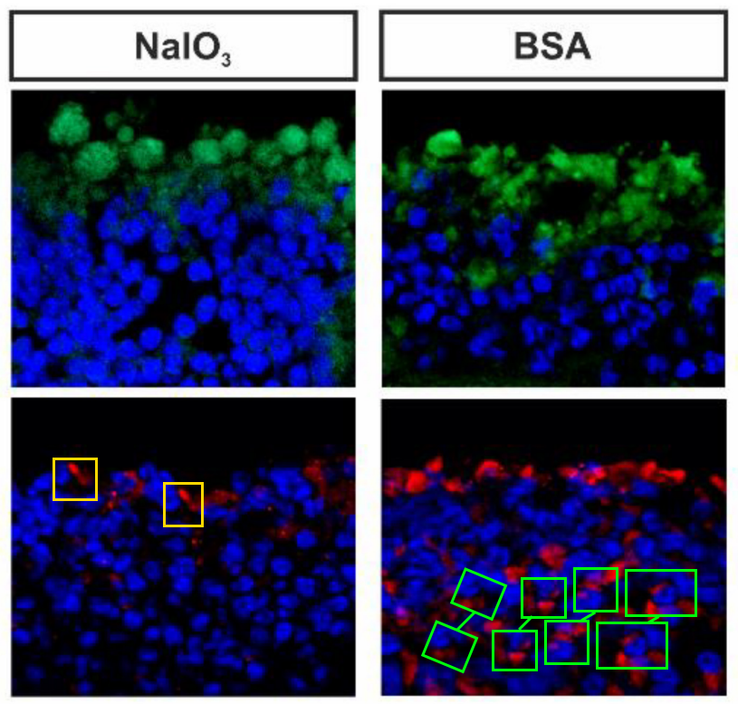
Figure 7A (again): Green shapes indicate the duplicated areas.
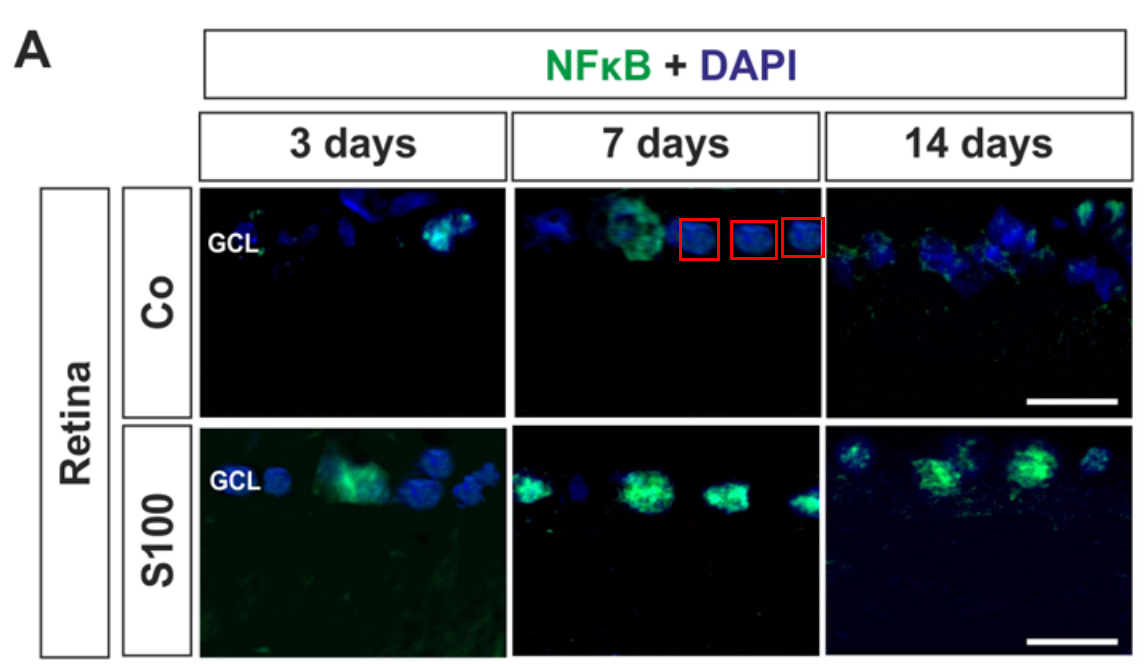
The third concern I described above (apart from the duplication and the cloning) is the manipulation of images with a paintbrush type tool. I suspect that a very large number of images have been adjusted in this way, but not all of them are easy to see. Here’s my best demonstration from the supplementary section of a 2018 Scientific Reports paper (where you will notice that Stephanie works with both Faissners, father and son). I should say this issue is easiest to see on a bright screen, especially a PC monitor.
Sabrina Reinehr, Jacqueline Reinhard, Marcel Gandej, Ivo Gottschalk, Gesa Stute, Andreas Faissner, H. Burkhard Dick, Stephanie C. Joachim S100B immunization triggers NFκB and complement activation in an autoimmune glaucoma model Scientific Reports (2018) doi: 10.1038/s41598-018-28183-6

Figure S3 from the same paper also appears to have been digitally painted on, only this time with a thicker brush setting. Again I have increased the brightness and contrast.
The effect is most obvious in the lower two images in this arrangement, although the upper right image also appears to have been painted on.
In Figure 4A of the same paper (below) the painting effect is harder to see, but there are also some cloned cells which should be quite obvious, I have added the red rectangles to show where I mean.
This fine art was sponsored by DFG, who just recently awarded Reinehr with a research grant. No wonder DFG provided Leonid and I with this (very) expert opinion about Reinehr’s and Joachim’s papers (translated):
“...according to the DFG Rules of Procedure for Dealing with Scientific Misconduct (§ 14), the DFG can only initiate proceedings if the reported suspicion is sufficiently concrete. The notification must clearly indicate which specific actions or which specific findings constitute scientific misconduct.
As a precautionary measure, we would like to point out that the notification of automatically identified similarities (“much more similar than expected”) does not generally meet this requirement. Such similarities initially only represent a formal consensus, but they do not contain any scientific justification or analysis as to why the consensus could constitute scientific misconduct.“
Here’s a 2021 paper from Frontiers in Cellular Neuroscience, funded by DFG, RUB and a grant from the Ernst and Berta Grimmke foundation to Reinehr:
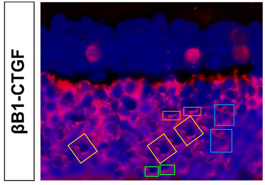
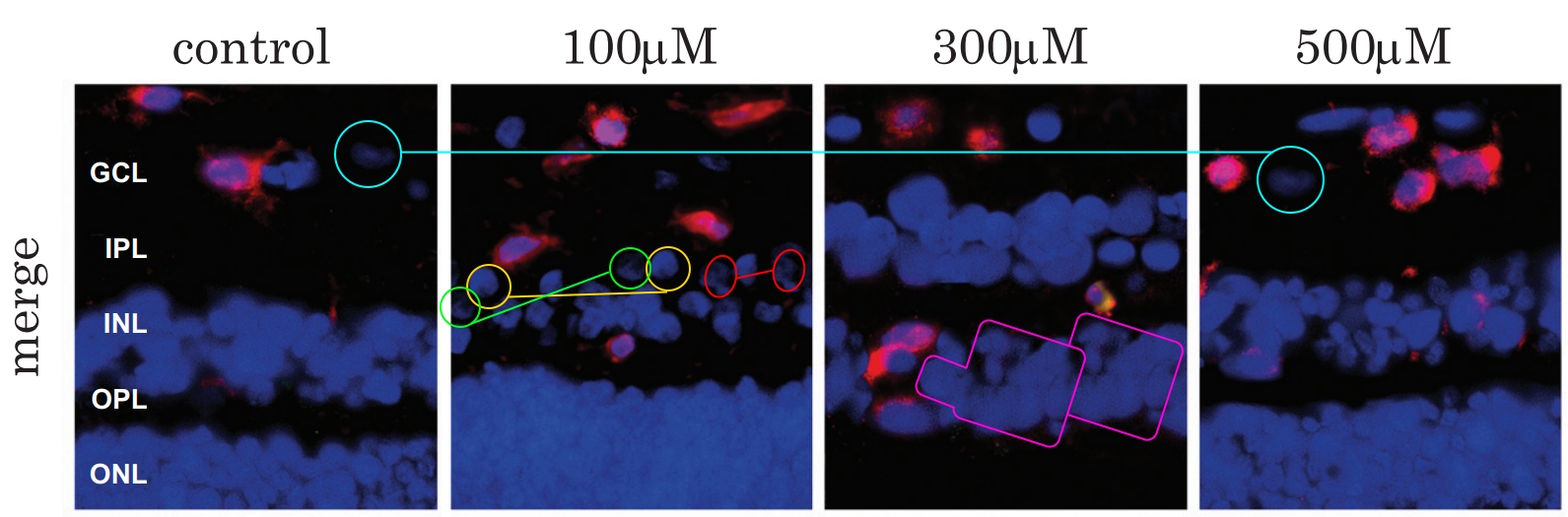
Sabrina Reinehr, Johanna D. Doerner, Ana M. Mueller-Buehl, Dennis Koch, Rudolf Fuchshofer, H. Burkhard Dick, Stephanie C. Joachim Cytokine and Complement Response in the Glaucomatous βB1-CTGF Mouse Model Frontiers in Cellular Neuroscience (2021) doi: 10.3389/fncel.2021.718087
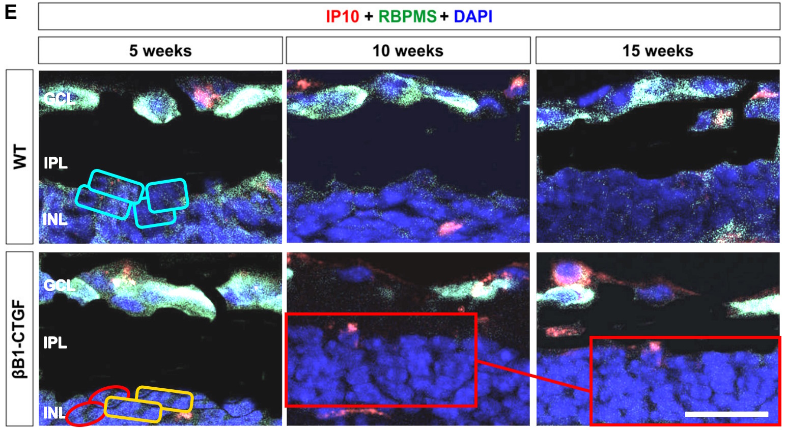
In Figure 6E you can see unnaturally dark background scribbles, cloned individual elements, and a large section of recycled DAPI staining where one of the images has been substantially modified with painting to make them appear different. Again, In this image I have increased the brightness and contrast to help visualise these elements. If you’re still struggling to see the scribbling hopefully the image from Figure 5 should be completely convincing:

There are plenty more problematic images in this paper.
Although some of these images have been fairly crudely edited, I would say almost all of the modifications pass the immediate smell test and many of them will also escape automated checking. Frankly, some of these have been done very well, and I strongly suspect I have missed many (if not most) of them.
Here’s one from Figure 5A of a paper in Journal of Cellular and Molecular Medicine, also funded by DFG and the Grimmke foundation:
Sabrina Reinehr, Dennis Koch, Maximilian Weiss, Franziska Froemel, Christina Voss, H. Burkhard Dick, Rudolf Fuchshofer, Stephanie C. Joachim Loss of retinal ganglion cells in a new genetic mouse model for primary open‐angle glaucoma Journal of Cellular and Molecular Medicine (2019) doi: 10.1111/jcmm.14433
I found no other images with cloned sections in this paper, but I suspect there must be more.
Unlike (for example) Egyptian histology where modifications seem to be introduced haphazardly with no specific goal (apart from perhaps evading the detection of duplicate images), I get the sense that the editing in Stephanie’s papers is carefully targeted towards individual areas of images and selected experimental conditions to strengthen the arguments being made in the paper.

Egyptian Toxicology Mortal Combat
“Stupid people do stupid things, After all it was an Egyptian who once told me: “10% editing is acceptable as long as we didn’t modify the significant ” – Sholto David
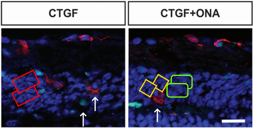
Here’s an example from another DFG-funded paper in Journal of Cellular and Molecular Medicine, Faissner Senior is also on board. The images with the red rectangles are derived from the same original and the modified area is exactly where the author’s white arrows are pointing. In this case the longer green extensions have been digitally ablated. Dare I say this would impact conclusions?
Sabrina Reinehr, Jacqueline Reinhard, Susanne Wiemann, Gesa Stute, Sandra Kuehn, Julia Woestmann, H. Burkhard Dick, Andreas Faissner, Stephanie C. Joachim Early remodelling of the extracellular matrix proteins tenascin‐C and phosphacan in retina and optic nerve of an experimental autoimmune glaucoma model Journal of Cellular and Molecular Medicine (2016) doi: 10.1111/jcmm.12909
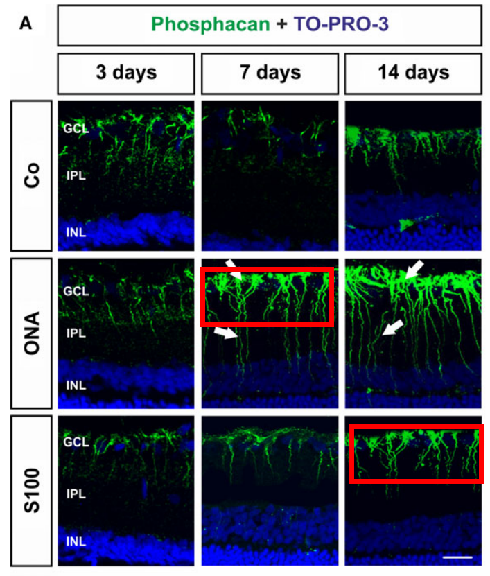
Figure 2A: Green signal (and DAPI) are highly similar (but not the same) between two images that should show different treatment conditions/time points.
Here’s another image with very precise modifications, I suspect these additional cells were added to make a more favourable comparison to the adjacent “Aged” samples (not shown below). The only funding for this study came from the FoRUM programme for medical research by RUB:
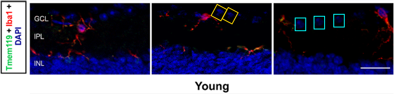
Clivia Erb, Sabrina Reinehr, Carsten Theiss, H. Burkhard Dick, Stephanie C. Joachim HSP27 induced glaucomatous damage in mice of young and advanced age Frontiers in Cellular Neuroscience (2023) doi: 10.3389/fncel.2023.1257297
Scientific image forgers make stylistic choices. Scribbling with the paintbrush tool and copy-move forgery with a soft edged tool are characteristic of whoever has been making these faked images. At first I wondered if Stephanie Joachim or Sabrina Reinehr was the artist in residence, as they are both authors on almost all of the seriously troubled papers. However, only Stephanie is an author of this comparatively early effort from 8 years ago. This indicates to me that it is Stephanie herself who should be asking difficult questions about research ethics.
José Hurst, Sandra Kuehn, Adelina Jashari, Teresa Tsai, Karl Ulrich Bartz-Schmidt, Sven Schnichels, Stephanie C. Joachim A Novel Porcine Ex Vivo Retina Culture Model for Oxidative Stress Induced by H2O2 Alternatives to laboratory animals (2017) doi: 10.1177/026119291704500105
There is also another large overlapping section in this paper, but without any manipulation with the clone or brush tool that I can see.
The study was funded by the German Federal Institute for Risk Assessment (BfR), and it was published in the journal Alternatives to laboratory animals. To be fair, fraud is certainly a substitute for using animals in the lab.
Here’s another paper where Sabrina did not participate. It was funded by Grimmke foundation, this time via a grant to Joachim’s former PhD student Sandra Kühn:
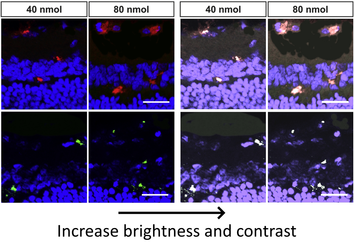
Pia Grotegut, Sandra Kuehn, Wilhelm Meißner, H Burkhard Dick, Stephanie C Joachim Intravitreal S100B Injection Triggers a Time-Dependent Microglia Response in a Pro-Inflammatory Manner in Retina and Optic Nerve Molecular Neurobiology (2020) doi: 10.1007/s12035-019-01786-4

Of course it is possible that more than one person in the lab has the habit of modifying mages, here’s an example from 2023 where some inserted areas have sharp edges, I’ve added the white arrows to show where I mean. There are also duplicate areas in the coloured shapes, and some darker scribbling with a narrower tool in the background. Funding provided by DFG and Glück im Blick:
Sabrina Reinehr, Renée M. Girbig, Kim K. Schulte, Janine Theile, M. Ali Asaad, Rudolf Fuchshofer, H. Burkhard Dick, Stephanie C. Joachim Enhanced glaucomatous damage accompanied by glial response in a new multifactorial mouse model Frontiers in Immunology (2023) doi: 10.3389/fimmu.2022.1017076
Here’s another one with sharp edges visible in the cloned areas, funded by RUB’s FoRUM program and the Hertie Foundation, which “promotes neuroscience and democracy” and was charged with embezzlement and tax evasion 25 years ago. Dick the Boss is there, and Faissner Junior is penultimate author:
Laura Petrikowski, Sabrina Reinehr, Steffen Haupeltshofer, Leonie Deppe, Florian Graz, Ingo Kleiter, H Burkhard Dick, Ralf Gold, Simon Faissner, Stephanie C Joachim Progressive Retinal and Optic Nerve Damage in a Mouse Model of Spontaneous Opticospinal Encephalomyelitis Frontiers in Immunology (2021) doi: 10.3389/fimmu.2021.759389

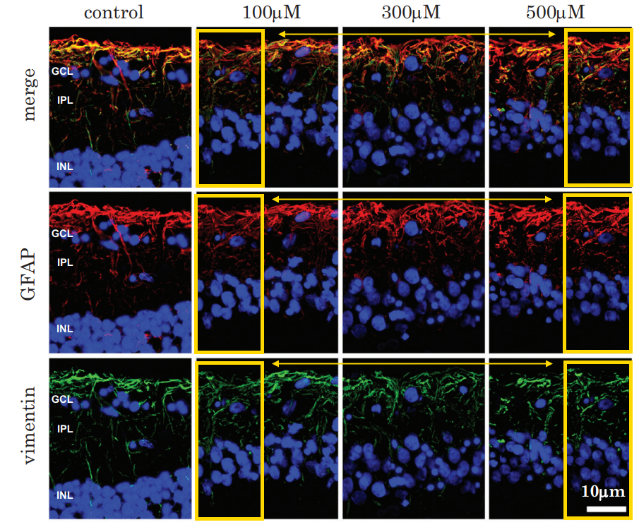
Although most of the forgeries have been conducted on immunofluorescence images, other types of images have been manipulated too. The German pharma giant Bayer sponsored this piece in Frontiers in Cellular Neuroscience in 2019:
Marina Palmhof, Viktoria Frank, Pascal Rappard, Emely Kortenhorn, Julia Demuth, Nora Biert, Gesa Stute, H Burkhard Dick, Stephanie C Joachim From Ganglion Cell to Photoreceptor Layer: Timeline of Deterioration in a Rat Ischemia/Reperfusion Model Frontiers in Cellular Neuroscience (2019) doi: 10.3389/fncel.2019.00174
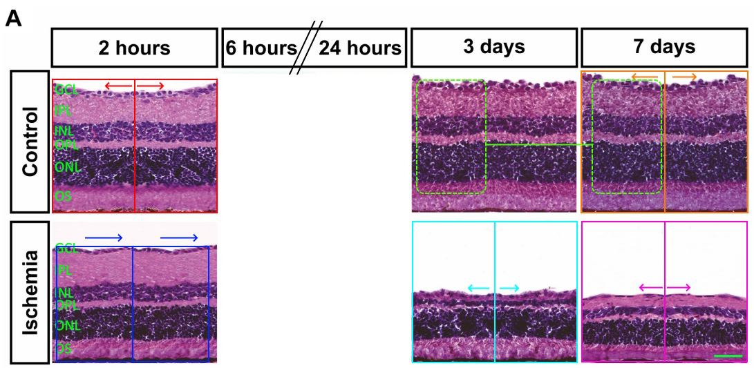
The light microscopy images of stained eyes have been enlarged by mirroring or duplicating large blocks through the centre of the image. These mirrored sections have a few differences too, so they must have also been doctored. The same paper includes too many edited images to show all of them, but here are some more scribbles and cloned areas of DAPI staining:

I’ll show one more example of scribbling, fortunately no one paid for this, the funding statement is emphatic: “This research did not receive any specific grants from funding agencies in the public, commercial, or not-for-profit sectors.” Normally, mouse research is quite expensive, I guess it was just out of the kindness of their hearts to contribute this for scientific community!
Sandra Kuehn, Sabrina Reinehr, Gesa Stute, Cara Rodust, Pia Grotegut, Alexander-Tobias Hensel, H. Burkhard Dick, Stephanie C. Joachim Interaction of complement system and microglia activation in retina and optic nerve in a NMDA damage model Molecular and Cellular Neuroscience (2018) doi: 10.1016/j.mcn.2018.05.001
This same paper has a more typical overlapping image, pay closer attention to the DAPI.

There are more papers where I only found boring types of overlaps, readers are encouraged to investigate… Sponsored by Grimmke Foundation and DOG, while DFG paid for open access publishing:
Pia Grotegut, Natarajan Perumal, Sandra Kuehn, Andreas Smit, H. Burkhard Dick, Franz H. Grus, Stephanie C. Joachim Minocycline reduces inflammatory response and cell death in a S100B retina degeneration model Journal of Neuroinflammation (2020) doi: 10.1186/s12974-020-02012-y

This study, sponsored by Bayer and RUB’s own Esser Foundation was even corrected one year after publication, simply to make it Open Access:
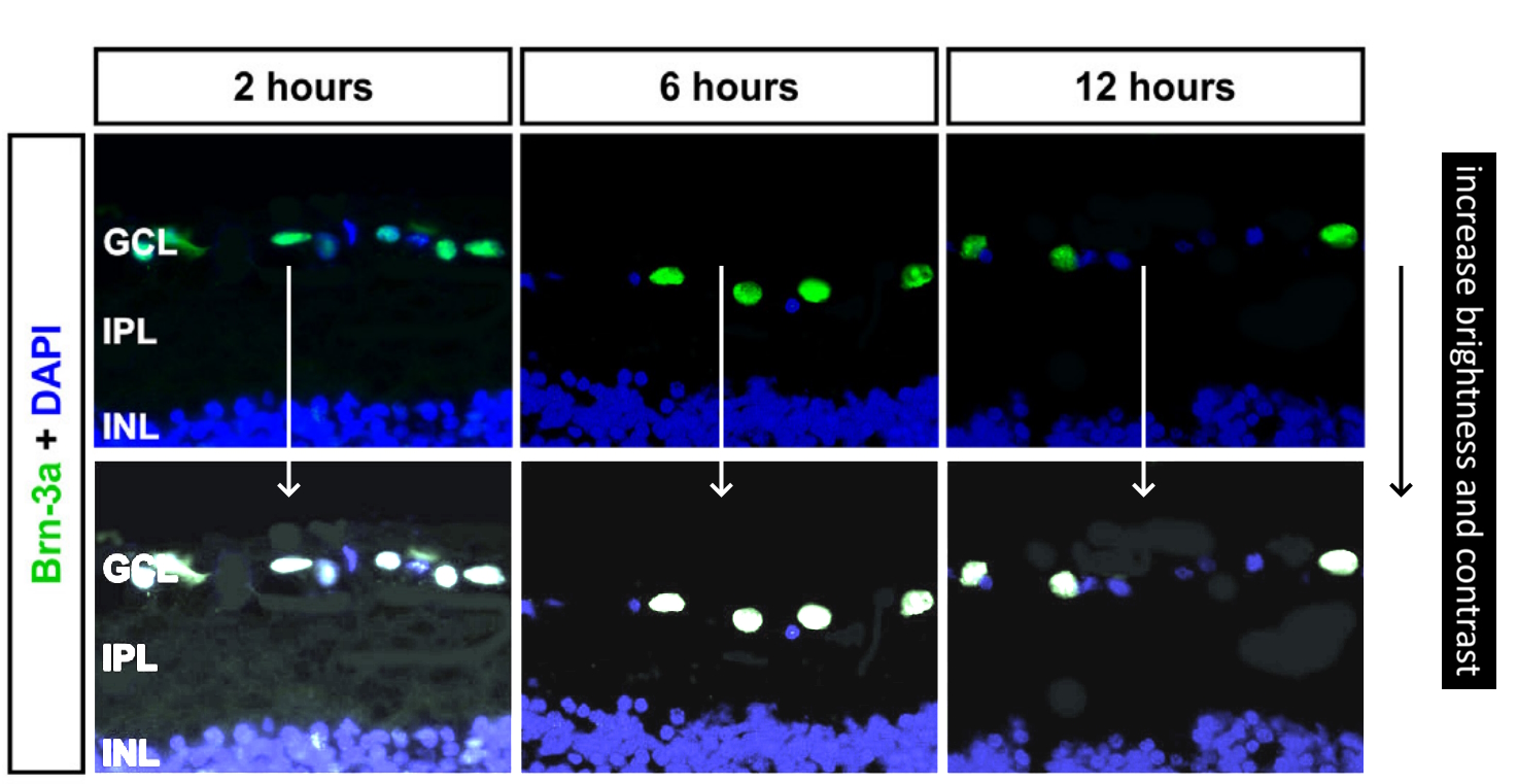
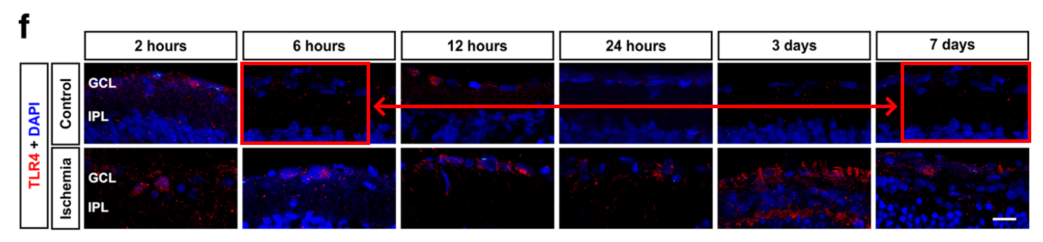
Natalie Wagner, Sabrina Reinehr, Marina Palmhof, David Schuschel, Teresa Tsai, Emely Sommer, Viktoria Frank, Gesa Stute, H. Burkhard Dick, Stephanie C. Joachim Microglia Activation in Retinal Ischemia Triggers Cytokine and Toll-Like Receptor Response Journal of Molecular Neuroscience (2020) doi: 10.1007/s12031-020-01674-w
This MDPI study, with Faissner Senior, was sponsored by DFG and the Konrad-Adenauer Stiftung, which is associated with the German party Christian Democratic Union (CDU).
Sabrina Reinehr, Jacqueline Reinhard, Susanne Wiemann, Karoline Hesse, Christina Voss, Marcel Gandej, H Burkhard Dick, Andreas Faissner, Stephanie C Joachim Transfer of the Experimental Autoimmune Glaucoma Model from Rats to Mice-New Options to Study Glaucoma Disease International Journal of Molecular Sciences (2019) doi: 10.3390/ijms20102563
There are also examples of image recycling between papers, see here for example, the experimental conditions do not appear to be the same for the yellow rectangles. Both studies were sponsored by the Pro Retina Foundation, the first one also by the pharma giant Novartis:
- Natalie Wagner, Armin Safaei, José Hurst, Pia A. Vogt, H. Burkhard Dick, Stephanie C. Joachim, Sven Schnichels Impact of Primary RPE Cells in a Porcine Organotypic Co-Cultivation Model Biomolecules (2022) doi: 10.3390/biom12070990
- Natalie Wagner, Armin Safaei, Pia A. Vogt, Maurice R. Gammel, H. Burkhard Dick, Sven Schnichels, Stephanie C. Joachim Coculture of ARPE-19 Cells and Porcine Neural Retina as an Ex Vivo Retinal Model Alternatives to laboratory animals (2022) doi: 10.1177/02611929221082662
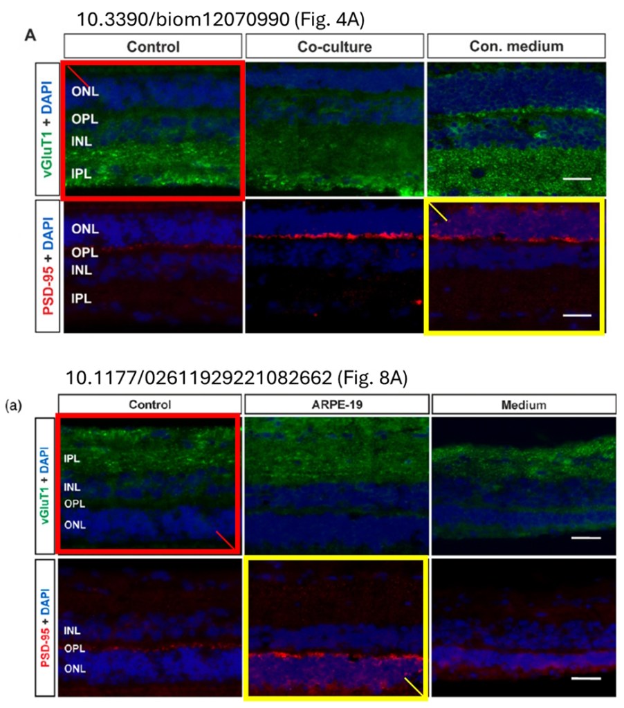
Curculio davidi: Two images in Fig.4A are identical to previously published images by the same group after a 180° rotation.
Joachim’s mentee Sven Schnichels is another Young DOG.
I mentioned earlier that none of the images in Stephanie’s papers have previously been criticised. However, there was certainly an opportunity to discover this disaster sooner.
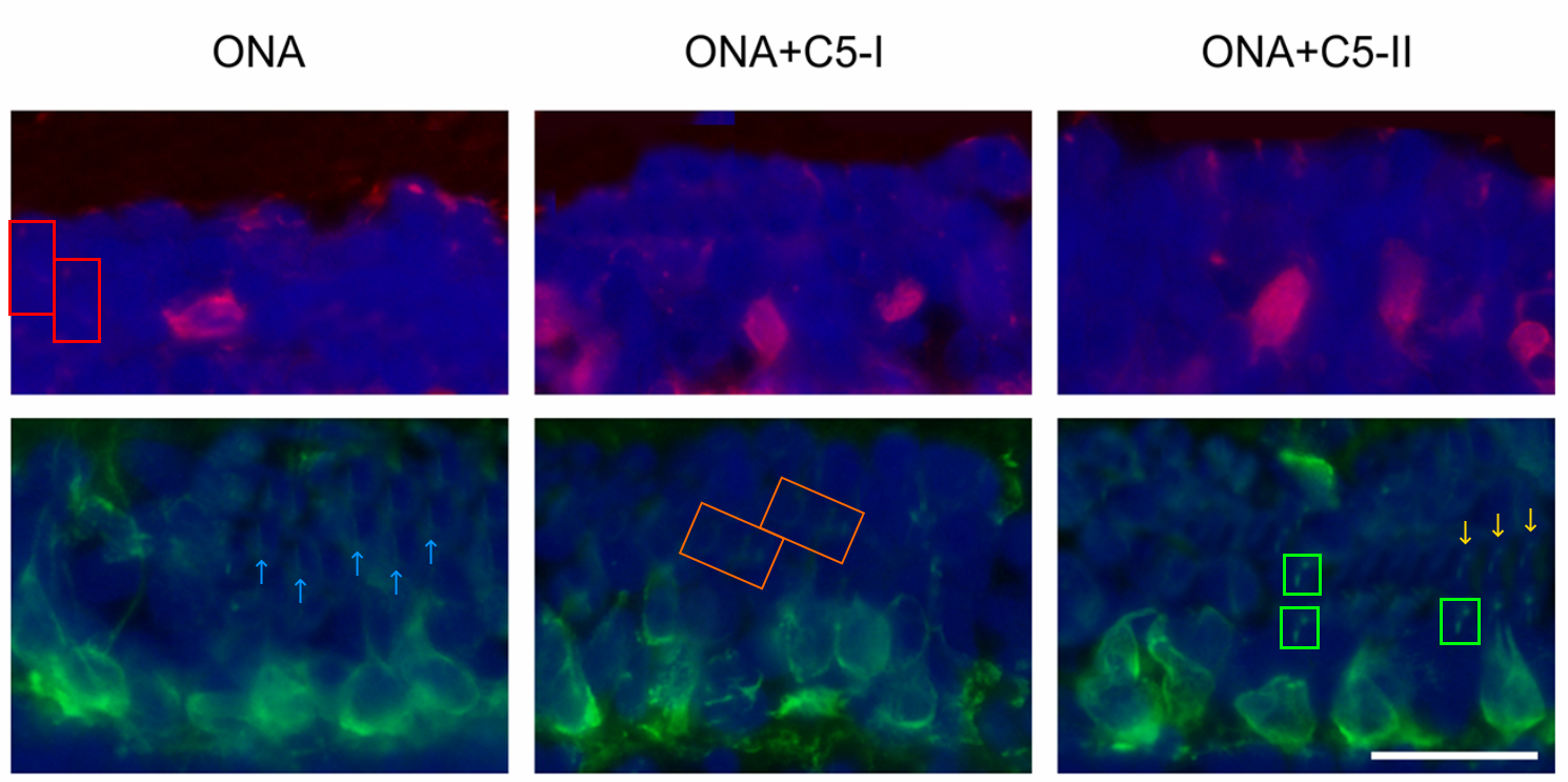
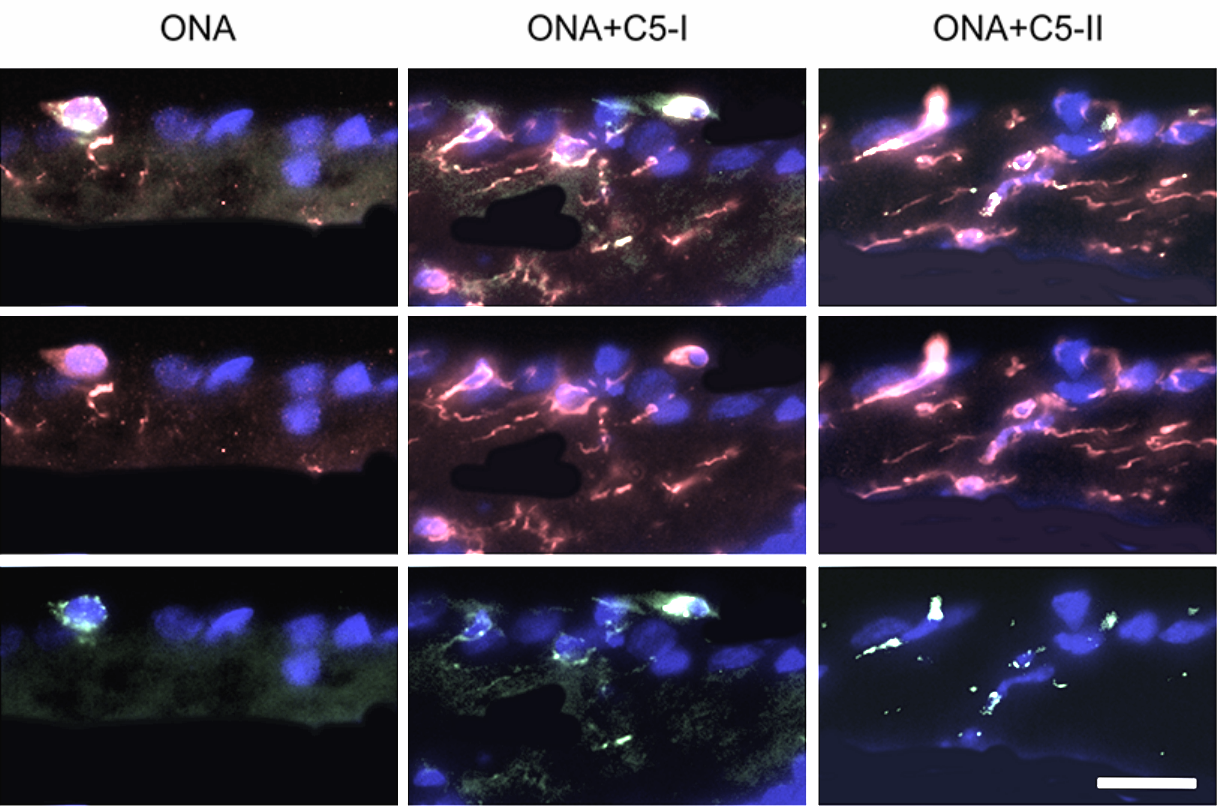
Sabrina Reinehr, Sara C. Gomes, Caroline J. Gassel, M. Ali Asaad, Gesa Stute, Marc Schargus, H. Burkhard Dick, Stephanie C. Joachim Intravitreal Therapy Against the Complement Factor C5 Prevents Retinal Degeneration in an Experimental Autoimmune Glaucoma Model Frontiers in Pharmacology (2019) doi: 10.3389/fphar.2019.01381
In 2022, an anonymous user wrote on PubPeer:
“Serious issue with the design of this study: BB5.1 antibody used here does not work in rats, though the entire study was done in rats. BB5.1 inhibits C5 only in mouse models and this can be easily found in BLA documents (Eculizumab).
Lutzomyia tortura
Second, how come strong conclusions are made, such as ” Our results indicate that the C5 antibody inhibits activation of the complement system, preventing the loss of retinal function”, however, most of the data presented are not statistically significant?“
The comment seems to be spot-on, see Zelek et al 2020 for example: “BB5.1 efficiently inhibited C5 in mouse serum but not in human or other rodent sera“. Certainly it deserves a response from the authors, but of course this comment was ignored. Had anyone bothered to look carefully, they would have discovered the whole thing is a sham, complete with silly scribbles and outrageously doctored images. Well, perhaps neither the experiments nor the images affect the conclusions? This was sponsored by DFG and a Grimmke Foundation grant to Reinehr.
Once again I am left with the impression that it is not lack of information that is preventing such fraud from being stopped, but lack of interest or curiosity, after all, there is a PubPeer dashboard for institutions and journals, don’t they care what people are saying about their research?
It seems hard to accept any explanation that doesn’t somehow incriminate most of the people involved, H. Burkhard Dick is an author on almost all of these papers, but has he ever read them, never mind examined the images? How are we to imagine the laboratory was functioning? In my own experience I have discussed the minutia of almost every data type in painful detail, and I don’t think I would’ve missed a giant black splodge on an image, especially if it was one that I had taken. I reached out to everyone involved and they all ignored me, so I reached out to every faculty member of the International Graduate School of Neuroscience (which Stephanie is a member of), and finally received a response (a handy tip there, for anyone struggling with people who don’t like to reply to emails).
Dear All,
Ulf Eysel (Ombudsstelle)
this matter was brought to the attention of the Ombudsstelle of the Ruhr-University on 26/27 October 2025 which has taken first action to clarify the evidence on 27 October 2025.
Best regards,

DFG decision: Antonia Joussen innocent victim of co-authors’ data manipulations
“The committee […] requests of you for future publications to assess well ahead and to question critically your responsibility for the contributions of the co-authors, also for your own protection.”
We can certainly say based on the evidence that research into blindness is important in Germany, so perhaps DFG should fund someone to find out why so many scientists are suffering from vision loss? But I can’t say I am filled with optimism. Expect DFG to find Joachim innocent (as they did for fellow blind(ness) researcher Antonia Joussen). Perhaps the ideal candidate to lead the exoneration investigation would be Professor Bernhard Sabel, Germany’s celebrity hero of research integrity. Maybe it’s no coincidence that also Sabel cures blindness:


They are blinding us with science: https://www.youtube.com/watch?v=V83JR2IoI8k
LikeLiked by 1 person
‘Dare I say this would impact conclusions?’
Of course not! Such image adjustments were made solely for aesthetic clarity and never alter the underlying data or conclusions, they do serve as a reminder, however, that beauty – particularly in ophthalmic research – remains in the eye of the beholder.
LikeLiked by 1 person
Fair point, clearly I still have a lot to learn about conclusions. Maybe with enough research one day we can discover a fact or observation that does affect the conclusions.
LikeLiked by 1 person
Now, that would be ‘unfair’, wouldn’t it?
LikeLike
It’s shocking how pervasive image manipulation seems to be, especially in fields that rely so heavily on visual data like ophthalmology
LikeLiked by 2 people
I remain perpetually surprised, to be honest.
LikeLike
Stockholm Declaration, Section #4, Item 4 reads:
”Reward sleuthing, but beware of unfair ‘whistle-blowers’.”
I was wondering what inspired the committee members such as Bernhard Sabel, the Science and Innovation Integrity Foundation (Sciii gGmbH) founder and Peter Seeberger the Vice President of DFG, senate member of the Max Planck Society to add the second part.
But the sentence feels incomplete, perhaps someone inadvertently left out the best part. Let me help to fill out the rest with some educated guess: ”Reward sleuthing, but beware of unfair ‘whiste-blowers’ and dismissive research integrity offices”.
LikeLiked by 1 person
Unfair whistleblowers is us, Sholto, me, Aneurus, Magazinov etc
The good sleuths are Byrne & Christopher and those in residence of Retraction Watch.
LikeLike
Being philistine is unfair indeed (to the world of art).
Good sleuths are neither philistine nor puerile. [I’m trying to practice my new vocabulary by using it in a sentence, if you don’t mind]
LikeLike
Individually adjusted photoshopping of scientific images is a holistic complement to traditional research such as doing laboratory experiments.
LikeLiked by 2 people
Amazing read. Curious to see how this story will be handled by RUB and DFG.
LikeLiked by 1 person
Frontiers have been on top of things these days, I’ll need to get in touch with them.
LikeLike
I think Sholto David is an extreme utilitarian who considers himself fair, yet in reality, he constantly targets ordinary researchers. If you really have the capability, why not scrutinize the papers of all the renowned academicians and experts in the UK, the US, and Germany—and maybe even establish an integrity index afterward? It seems as though Indians or Chinese have offended you personally, given how exclusively you focus on them. That kind of behavior is the height of utilitarianism. It is also alleged that you accept payment to specifically debunk certain individuals—such conduct is truly unethical.
LikeLike
You do know you commented on an article about German professors?
LikeLiked by 1 person