
In my previous article, I presented the image integrity concerns voiced by Elisabeth Bik regarding the publications by the German medical diagnostics company Euroimmun. It was about a mysteriously photoshopped image of an assay chip as well as some possibly more serious assay data duplications, which the collaborating academic institutions, the University of Lübeck and the Robert-Koch-Institute announced to investigate.
EuroImmun was bought for $1.3 Billion by the US biotech giant PerkinElmer in 2017, but the new owner chose never to reply to my messages. Instead, Euroimmun’s founder and former CEO Winfried Stöcker provided a point-by-point reply on every criticised paper. The replies were co-signed by Euroimmun’s new CEO Wolfgang Schlumberger, both gentlemen are also key authors on the criticised publications.
The photoshopped chips’ mystery did however not become clearer even after Stöcker’s reply. Euroimmun’s sales director Kai Fechner previously educated me that the chips are not research data, but graphic art. Which still did not explain why going to such lengths to photoshop a chip picture, with new wells and new labelling, instead of simply snapping a photo of a proper chip with your smartphone. Stöcker’s reply is even more confusing. He informs us that
“In some publications, microscopic pictures mounted into Euroimmun slides were shown. The photos were not directly taken from an incubated slide itself, this is technically not possible. They are taken using a fluorescence microscope and inserted at the computer into the document – for easier understanding into a mask which looks like an Euroimmun slide. To investigate into the fingerprint of the plastic slide is really nonsense. The essential message of this publication is beyond any question.”
Ah. Who knew. Thank you Professor Stöcker, we learned something about microscopy, not just yet clear what. But then again, the chip image is not decorated with tiny pictures of fluorescent assays taken with microscope. The figures simply show a photoshopped chip, which returns again and again with differently shaped wells and different labelling. Stöcker’s reply still does not explain the need for the graphic art efforts.
Stöcker and Schlumberger sum up:
“In summary, Dr. Elisabeth Bik’s observations are not suitable to bring us into disrepute. She will not find a more integer company as Euroimmun, where enthusiastic scientists considerably contribute to the world’s technical and scientific progress since 40 years, including the period before starting the business. Thousands of people owe their lifes or the recovery of their health to our efforts and competence. And three thousand coworkers are proud to work at EUROIMMUN.”
I am not sure this disrepute bringing was what Bik tried to do, she rather wanted to help Euroimmun spot mistakes and improve their quality control, because it is not just the images of the chip which were found duplicated, but also actual assay data. Stöcker seems to agree that the fluorescence images are duplicated but sees the mistakes as unintentional. Asked about possible literature corrections, he said:
“Given the insignificance, we will inform the journals, but suggest to leave the matter rest“.
Following are the PubPeer evidence and comments posted by Bik, now supplemented with the explanations provided by Stöcker and Schlumberger.
Waltraud Suer, Cornelia Dähnrich, Wolfgang Schlumberger, Winfried Stöcker Autoantibodies in SLE but not in scleroderma react with protein-stripped nucleosomes Journal of Autoimmunity (2004) 10.1016/j.jaut.2004.02.002
Somebody apparently placed a dark rectangle over a part of the last lane, a rather unconventional practice of gel data presentation. Bik observed:
“In the “Nu2 (native)” lane, a completely blank (no background) area appears to be visible in the very relevant “Nucleosomal DNA” area. Image made a bit lighter to bring out the background. Shown with a thin red box.”

Stöcker explains:
“We were able to clearly discriminate systemic lupus erythematodes from progressive systemic sclerosis after we have observed that earlier nucleosome preparations were contaminated by DNA topoisomerase due to the affinity of this enzyme to its substrate. A very important discovery which excluded an assumed clinical overlap.
One of the chromatographic lanes demonstrated by Mrs. Bik really looks like to be covered at the lower end. We use to read each of our papers very carefully before being published, but did not observe such irregularity and cannot more find out anymore who caused it, we or the publisher. We would not have had any reason to improve the picture, since this separation is very easy: In the many years after this publication until now, for reagent production, we use the same procedure to separate nucleosomes from DNA topoisomerase, and the chromatographic quality control always provides the same clear result, the lower end of the chromatogram is always clean. At least, the essential message of this publication is beyond any question.”
It seems the publisher Elsevier resorted to secretly manipulating research data of unsuspecting authors, e.g. by placing dark boxes over parts of the gel track at exactly same position where undesired nucleosomal DNA might or might not have appeared. Good that none of that affected the essential message!
Karen Sonnenberg, Matthias Niedrig , Katja Steinhagen , Edda Rohwäder , Wolfgang Meyer , Wolfgang Schlumberger, Ewald Müller-Kunert , Winfried Stöcker State-of-the-art serological techniques for detection of antibodies against tick-borne encephalitis virus Int. J. Med.Microbiol. (2004) doi: 10.1016/s1433-1128(04)80028-7
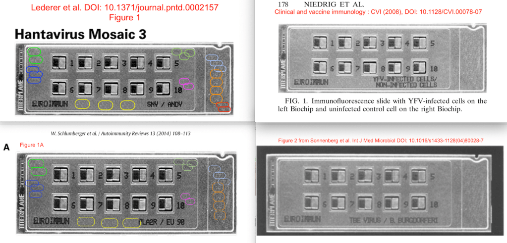
This is the first publication the “graphic art” chip made its appearance in. Bik described the problem:
- The top BIOCHIP slide (TBE virus / B. burgdorferi), showing 10 slots with 2 mosaic tiles each bears a striking resemblance to several other EuroImmun slides shown in other papers (DOI: 10.1128/CVI.00078-07, DOI: 10.1371/journal.pntd.0002157, DOI: 10.1016/j.autrev.2013.09.005, DOI: 10.3389/fimmu.2015.00221).
- In particular, Figure’s 2 top slide looks like the bottom slide (Hantavirus Mosaic 3) of Figure 1 of Lederer et al. PLOS NTD (2013), DOI: 10.1371/journal.pntd.0002157. Here is a comparison of the 2 figures with the similar-looking slides marked in aqua boxes.
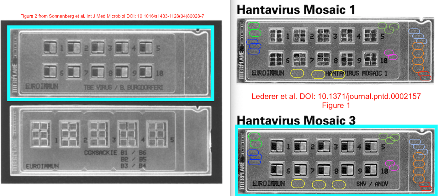
- In addition, these slides appear to show repetitive elements around the center mosaic tiles, as also found in several other papers by this group. These repetitive elements are shown in the Lederer figure above, in thin rounded boxes of the same color.”
Stöcker explains that the Sonnenberg et al paper:
“Describes the application of two elegant techniques (biochip-mosaics and line blots), incubated using titerplane technique, to diagnose tickborne encephalitis.
The photos are not taken from an incubated slide itself, this is technically not possible. They are taken using a fluorescence microscope and inserted at the computer into the document – for easier understanding into a mask which looks like an Euroimmun slide. To investigate into the fingerprint of the plastic slide is really nonsense. The essential message of this publication is beyond any question.”
EuroImmun’s encephalitis virus detection chip returned as Yellow Fever Virus (YFV) chip in 2008:
Matthias Niedrig , Oliver Kursteiner , Christian Herzog , Karen Sonnenberg Evaluation of an Indirect Immunofluorescence Assay for Detection of Immunoglobulin M (IgM) and IgG Antibodies against Yellow Fever Virus Clinical and Vaccine Immunology (2008) doi: 10.1128/CVI.00078-07
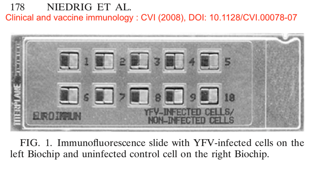
The first author is Matthias Niedrig, former head of the European Network for Imported Viral Diseases and professor at Robert-Koch-Institut, Germany’s top national authority on infectious diseases. Stöcker commented that this paper
“Uses biochip-duetts for the diagnosis of yellow fever. The photos are not taken from an incubated slide itself, this is technically not possible. They are taken using a fluorescence microscope and inserted at the computer into the document – for easier understanding into a mask which looks like an Euroimmun slide. To investigate into the fingerprint of the plastic slide is really nonsense. The essential message of this publication is beyond any question.”
In 2013, a very similar looking chip was used to detect hantavirus. Niedrig was now featuring as the last author:
Sabine Lederer , Erik Lattwein , Merle Hanke , Karen Sonnenberg , Winfried Stoecker , Åke Lundkvist , Antti Vaheri , Olli Vapalahti , Paul K. S. Chan , Heinz Feldmann , Daryl Dick , Jonas Schmidt-Chanasit , Paula Padula , Pablo A. Vial , Raluca Panculescu-Gatej , Cornelia Ceianu , Paul Heyman , Tatjana Avšič-Županc , Matthias Niedrig Indirect Immunofluorescence Assay for the Simultaneous Detection of Antibodies against Clinically Important Old and New World Hantaviruses PLoS Negl Trop Dis (2013) doi: 10.1371/journal.pntd.0002157
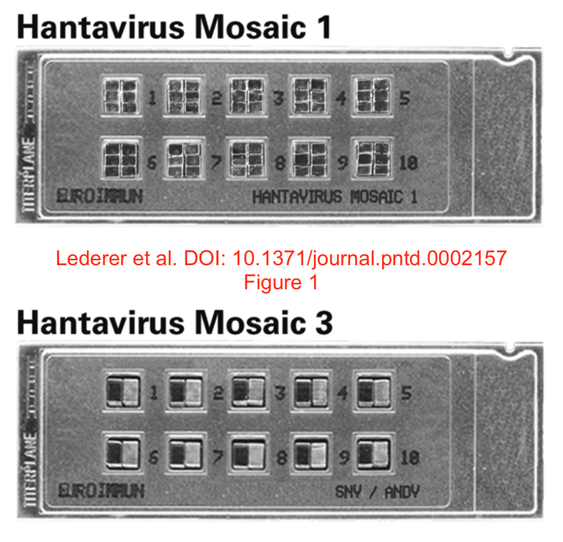
Bik noticed that also these chips ” show the same repetitive patterns”. Stöcker however says that Lederer et al paper
“Uses biochip-mosaics for the diagnosis and differentiation of Hanta virus infections. The photos are not taken from an incubated slide itself, this is technically not possible. They are taken using a fluorescence microscope and inserted at the computer into the document – for easier understanding into a mask which looks like an Euroimmun slide. To investigate into the fingerprint of the plastic slide is really nonsense. The essential message of this publication is beyond any question.”
In 2014, the chip returned to prance around in a review, authored by both Stöcker and Schlumberger. Now the chip was used for autoimmune diseases, instead of viruses.
Wolfgang Schlumberger, Nora Hornig, Sascha Lange, Christian Probst, Lars Komorowski, Kai Fechner, Cornelia Dähnrich, Winfried Stöcker Differential diagnosis of membranous nephropathy with autoantibodies to phospholipase A2 receptor 1 Autoimmunity Reviews (2014) doi: 10.1016/j.autrev.2013.09.005
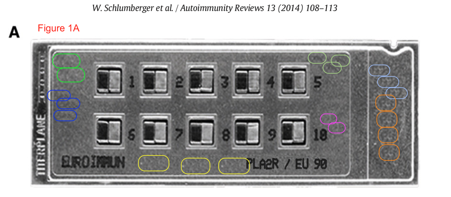
Here, Stöcker again explains that the paper
“Uses biochip-duetts for the diagnosis of membraneous nephropathy by detection of autoantibocies aganist phospholipase-A2 receptors 1.
The photos are not taken from an incubated slide itself, this is technically not possible. They are taken using a fluorescence microscope and inserted at the computer into the document – for easier understanding into a mask which looks like an Euroimmun slide. To investigate into the fingerprint of the plastic slide is really nonsense. The essential message of this publication is beyond any question.”
Bik is cannot follow this argument, but she merely did her PhD in microbiology and worked in this field of research for decades. She told me:
“I disagree that these are “obviously” photographic composites, and I find the answers a bit dismissive. Any manufacturer of a product should know better than photoshopping their products, in particular in scientific papers.“
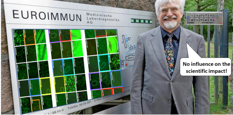
This was why on 24 January Stöcker clarified why the photoshopped chip makes perfect sense scientifically:
“In other publications, Euroimmun plastic slides were shown, equipped with biochips before incubation. Also here, the biochips were inserted virtually to demonstrate how these slides are configured, since most scientists are not acquainted with the biochip technology. Sonnenberg describes the application of two elegant techniques (biochip-mosaics and line blots), incubated using titerplane technique, to diagnose tick borne encephalitis. Niedrig uses biochip-duets for the diagnosis of yellow fever. Lederer uses biochip-mosaics for the diagnosis and differentiation of Hanta virus infections. Schlumberger uses biochip-duets for the diagnosis of membranous nephropathy by detection of autoantibodies against phospholipase-A2 receptors 1. All these publications contribute to the diagnostic progress, if Dr. Bik detects some irregularities, these at least did not influence the scientific message.”
It is strange. Do the real chips look so fearsome and mind-bogglingly disturbing that it would drive “most scientists” to insanity, were they to glimpse a picture of the actual chip used? Is that chip a diagnostics equivalent to the world’s deadliest joke where every Euroimmun employee was only allowed to see small bits of it, with the final image assembled in Photoshop?
Wenn ist das Nunstück git und Slotermeyer? Ja! Beiherhund das Oder die Flipperwaldt gersput!
In 2015, the world’s funniest chip finally made it into the world’s bestest (and the most ethical) journal family, Frontiers.
Gianna Mastroianni-Kirsztajn, Nora Hornig and Wolfgang Schlumberger Autoantibodies in renal diseases – clinical significance and recent developments in serological detection Frontiers in Immunology (2015) doi: 10.3389/fimmu.2015.00221
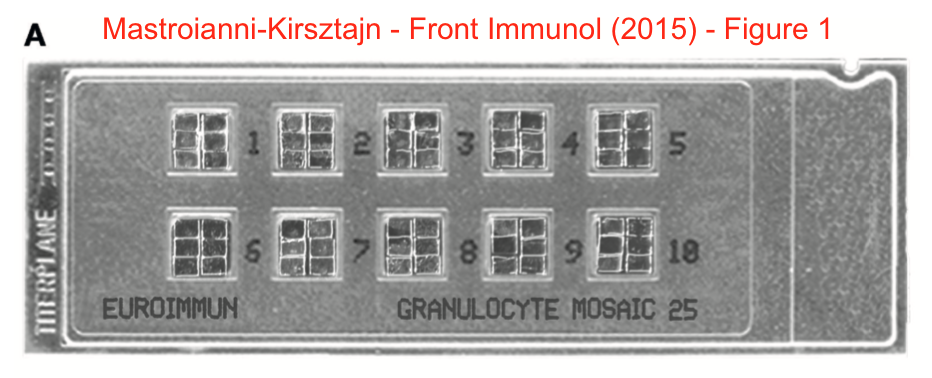
There was more: glomerular basement membrane (GBM) and myeloperoxidase (MPO) produced exactly same signal as EuroImmun’s proprietary indirect immunofluorescence microdots. Only rotated.
Stöcker says it doesn’t matter, since this Frontiers paper
“Describes a new very effective biochip mosaic tool to diagnose autoimmune bullous diseases by indirect immunofluorescence using a panel of different substrates.
The biochips used containing GBM microdots and MPO microdots are different. And even if not so, the essential message of the publication were not influenced and beyond any question, since the figure only describes a technique for better understanding. Not any calculation derives from this picture.”
The highlighted bit was added by Stöcker on 24.01.2020.
But what was the point of adding any research data to Euroimmun’s papers then if the essential message stands even without data? The next paper featuring Professor Stöcker had duplications in the actual chip assay:
Nina Van Beek , Kristin Rentzsch , Christian Probst , Lars Komorowski , Michael Kasperkiewicz , Kai Fechner , Inga M. Blöcker , Detlef Zillikens , Winfried Stöcker , Enno Schmidt Serological diagnosis of autoimmune bullous skin diseases: Prospective comparison of the BIOCHIP mosaic-based indirect immunofluorescence technique with the conventional multi-step single test strategy Orphanet Journal of Rare Diseases (2012) doi: 10.1186/1750-1172-7-49
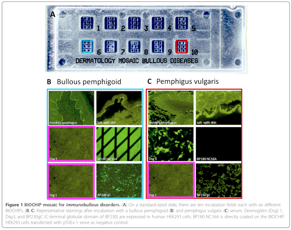
Here the authors highlighted two windows of the chip (Nr 6 and 10), and showed the staining on these in Figure 1B and 1C, respectively. Bik wrote:
“three of the 12 rectangles look very similar to each other. Shown with bright pink boxes. The teal and red boxes are part of the original figure. Image made lighter to bring out details. Note that the left Dsg 3 panel shows a couple of light spots not visible in the Dsg 1 panels.“
The last author Enno Schmidt is professor at, no pun intended of course, LIED. It is the institute for experimental dermatology at the University of Lübeck., the university announced to investigate the concerns. Stöcker explains (highlighted section added on 24.01.2020) that this Schmidt paper
“Compares indirect immunofluorescence using a powerful biochip mosaic with a conventional single test strategy.
Figure 1 is intended to explain the principle of the biochip mosaic technic, and not to evaluate samples. Obviously, the 3 pink surrounded pictures are identical. They show negative reactions. We should have used different substrates. We did not realize this mistake when supervising the manuscript. But it has not any influence on the scientific impact. Also here, the essential message of the publication were not influenced and beyond any question, since the figure only describes a technique for better understanding. Not any calculation derives from this picture.“
As reminder, Stöcker also does not see any real need to replace these images by a correction. Not even in the next case:
Christian Probst, Sandra Saschenbrecker, Winfried Stoecker, Lars Komorowski Anti-neuronal autoantibodies: Current diagnostic challenges Multiple Sclerosis and Related Disorders (2014) doi: 10.1016/j.msard.2013.12.001
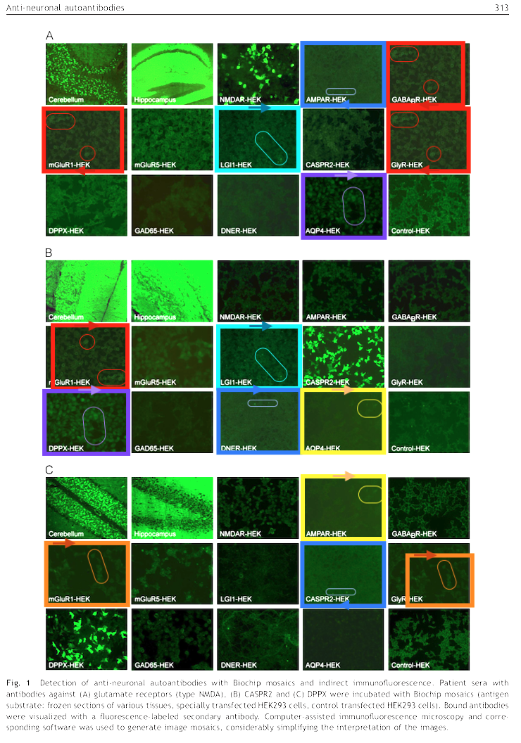
Last author Lars Komorowski is Euroimmun employee, but academically affiliated with the University of Lübeck (Stöcker himself holds an adjunct professorship at Lübeck). Bik raised serious concerns:
“As shown with rectangles of the same color, some of the panels look very similar to each other, albeit sometimes zoomed in differently, or rotated.”
Not so serious for Stöcker, it seems eminence trumps evidence any time. Here is what Stöcker says, brace yourselves:
“The authors belong to a group of the most successful scientists and clinical pathologists in the field of neuroimmunology worldwide, within the company Euroimmun. In this paper, their diagnostic techniques are described in principle.
Figure 1 is intended to explain the biochip mosaic technique, and not to evaluate samples. Obviously, to demonstrate several negative reactions, identical pictures have been used. It is a mistake which we did not realize when supervising the manuscript. But this carelessness has not any influence on the scientific impact.”
Ok, maybe inside Euroimmun the four authors are really “the most successful scientists and clinical pathologists in the field of neuroimmunology worldwide“, but outside of the Euroimmun walls? I really don’t know, are Probst, Saschenbrecker and Komorowski really such international giants that their research is 100% trustworthy even without the actual data? Also, how does one reuse image with different brightness, orientation and cropping, all by carelessness? Finally, the gigantic scientific impact of that paper is measured in exactly 9 PubMed-listed citations, of which 3 are coauthored by Euroimmun.
Bik is also puzzled by such dismissive attitude of the Euroimmun founder:
“[Stöcker] writes “figure 1 is intended to explain the principle of the biochip mosaic technique, and not to evaluate samples. Obviously, to demonstrate several negative reactions, identical pictures have been used.” This is remarkable, because the photos are presented as data.“
Cornelia Dähnrich , Lars Komorowski , Christian Probst , Barbara Seitz-Polski , Vincent Esnault , Jack F. Wetzels , Julia M. Hofstra , Elion Hoxha , Rolf A. Stahl , Gérard Lambeau , Winfried Stöcker , Wolfgang Schlumberger Development of a standardized ELISA for the determination of autoantibodies against human M-type phospholipase A2 receptor in primary membranous nephropathy Clinica Chimica Acta (2013) doi: 10.1016/j.cca.2013.03.015
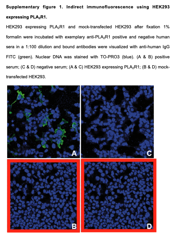
Bik noticed in the Figure S1. that
“Panels B (positive serum on mock transfected HEK293) and D (positive serum on mock transfected HEK293) appear to look very similar.”
Stöcker agrees that the cell image is duplicated and explains:
“The authors describe a standardized ELISA for the detection of autoantibodies against PLA2R to diagnose primary membranous nephropathy. A very powerful test system now used in hundreds of laboratories worldwide.
Also here, figure 1 is intended to explain the principle of the biochip mosaic technic, and not to evaluate samples. Obviously, to demonstrate several negative reactions, identical pictures have been erroneously inserted, without any influence on the scientific impact.”
Georgios Efthymiou, Efthymios Dardiotis, Christos Liaskos, Emmanouela Marou, Vana Tsimourtou, Thomas Scheper, Wolfgang Meyer, Alexandros Daponte, Lazaros I. Sakkas, Georgios Hadjigeorgiou, Dimitrios P. Bogdanos Anti-hsp60 antibody responses based on Helicobacter pylori in patients with multiple sclerosis: (ir)relevance to disease pathogenesis Journal of Neuroimmunology (2016) doi: 10.1016/j.jneuroim.2016.06.009
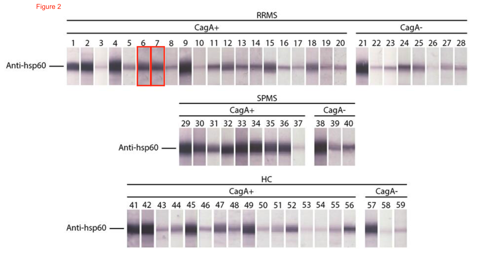
Here, Euroimmun only collaborated, their contributing authors are the two Germans in the middle of the Greek authors list, position 6 and 7. Just like on the gel in Figure 2: Thomas Scheper and Thomas Scheper. Or maybe it was Wolfgang Meyer and Wolfgang Meyer? Pardon the lame joke, very unprofessional of me, especially to repeat it. And also, Stöcker disagrees:
“Samples 6 and 7 resemble each other closely, but this may happen.”
I wonder under which circumstances this similarity may happen. Excessive carelessness? Here another collaboration, with Spanish partners this time:
Amilcar Perez-Riverol , Luís Gustavo Romani Fernandes , Alexis Musacchio Lasa , José Roberto Aparecido Dos Santos-Pinto , Débora Moitinho Abram , Gabriel Hideki Izuka Moraes , Frederic Jabs , Michaela Miehe, Henning Seismman , Mario Sergio Palma , Ricardo De Lima Zollner , Edzard Spillner , Márcia Regina Brochetto-Braga Phospholipase A1-based cross-reactivity among venoms of clinically relevant Hymenoptera from Neotropical and temperate regions Molecular Immunology (2018) doi: 10.1016/j.molimm.2017.11.007

Stöcker says here also:
“Two samples resemble each other closely, but this may happen as well.”
That is the strangest reply possible in such case. At least Stöcker could have said where the gels were made, so the authors can check their raw data, given that both papers from 2016 and 2018 are not really that ancient. Also Bik is disappointed:
“I had hoped [Stöcker] would have added the original figures. Just saying that they look similar but that “may happen” is not really a good answer. These are recent papers, so could he please show the original, high resolution/uncropped blots?“
This article was updated on 24.01.2020, to include Stöcker’s additional statements.
Update 31.01.2020
More Euroimmun assay image reuse, reported by Elisabeth Bik on PubPeer:
Myriam Schumacher , Frank Risto Rommel , Borros Arneth , Harald Renz , Winfried Stöcker , Anita Windhorst , Andreas Hahn , Bernd Axel Neubauer Encephalopathy Associated With Neurochondrin Autoantibodies Journal of Child Neurology (2019) doi: 10.1177/0883073819849773
“In this paper, three patients are presented with “autoimmune encephalitis”, and the patients’ serum was tested for the presence of autoantibodies against neurochondrin using indirect fluorescence with EuroImmun reagents (Figure 3). One author on this paper is/was an EuroImmun employee.
However, Figure 3 of this paper shows unexpected similarities to Figure 4 of a 2017 paper describing a different set of patients, but also EuroImmun assays. This paper is”:
R Miske , CC. Gross , M Scharf , KS. Golombeck , M Hartwig , U Bhatia , A Schulte-Mecklenbeck , K Bönte , C Strippel , L Schöls , M Synofzik , H Lohmann , I Madeleine Dettmann , M Deppe , S Mindorf , T Warnecke , Y Denno , B Teegen , C Probst , S Brakopp , KP Wandinger, H Wiendl, W Stöcker, SG. Meuth, L Komorowski, N Melzer Neurochondrin is a neuronal target antigen in autoimmune cerebellar degeneration Neurology(R) neuroimmunology & neuroinflammation (2017) doi: 10.1212/nxi.0000000000000307

“The 2017 paper (Miske et al.) describes three patients recruited at the Department of Neurology, University of Münster, Germany, with pronounced cerebellar and brainstem syndrome of unknown origin.
- Patient 1, male, 21 months history beginning at age 51y
- Patient 2, male, 54 months history beginning at age 23y
- Patient 3, female, 120 months history beginning at age 18y
This 2019 paper (Schumacher et al.) describes three patients recruited at the Department for Child Neurology at University hospital Gießen, with autoimmune encephalitis.
- Patient 1, male, age 6y
- Patient 2, female, age 7y
- Patient 3, male, age 14y
Figure 4 of the 2017 Miske et al. paper looks unexpectedly similar to Figure 3 of the 2019 Schumacher et al. paper (this paper). Shown with red boxes.“
The only common author on these two papers is Euroimmun founder Stöcker, he is the only one from Euroimmun on the 2019 paper from the University of Münster. On the 2017 paper from the University of Giessen, 10 authors are Euroimmun-affiliated. I will ask Professor Stöcker if he made a mistake when forwarding a wrong assay data to his Münster collaborators.

Donate!
If you are interested to support my work, you can leave here a small tip of $5. Or several of small tips, just increase the amount as you like (2x=€10; 5x=€25). Your generous patronage of my journalism will be most appreciated!
€5.00


Pingback: Euroimmun: Graphic Art, Made in Germany – For Better Science
It is a red flag when someone self-proclaimed to be the best in the world in any particular field of research….
LikeLiked by 1 person
When I saw Fig 1 I had a pretty hearty laugh. Guess they didn’t want to see any nucleosomal DNA in that mononuclosome prep.
LikeLike
Pingback: How David Ojcius and Jie Yan solved Leptospirosis – For Better Science
Pingback: German antivaxxers queue for Stöcker vaccine – For Better Science