On 13 December 2019, the American Chemical Society (ACS) issued a correction for a 3-year-old fraudulent paper. This correction is a representative declaration of bankruptcy for individual scholars, scholarly societies and scholarly publishing. If you were thinking of quitting science in disgust, this correction notice, and the following post by Smut Clyde might motivate you further to leave this fraud circus to professionals. Read at your own peril.
Not all is lost though. Even if our science elites beat their chests about research integrity while simultaneously peddling fraud and protecting fraudsters because these are their academic peers, we can at least point finger and laugh at them.
The corrected paper was Subramani et al ACS Applied Materials & Interfaces 2016, stemming from the lab of Dhanaraj Gopi, chemistry professor at Periyar University in India. Officially, Gopi does research in “Nanobiomaterials, Analytical Chemistry, Material Science and Corrosion”, in reality he and his lab mostly fake data in Photoshop on a good day, and on a bad day, they just draw some spectra with a pencil. That all was revealed in Gopi’s papers, including in the one ACS decided to correct instead of retracting. Figures containing fake images of cells cultured on even faker pie-nanocrusts were replaced with something less obviously fake, but even ACS didn’t dare correct a figure of alleged XRD spectra which were most obviously hand-drawn with a pencil or maybe with some coloured felt-tip markers. It passed peer review, so it’s science now, you see.

There are presently 32 Gopi papers discussed on PubPeer, one more fraudulent than the other. Smut Clyde addresses 24 of them in his guest post below. None of that will likely change ACS’ view on who the real troublemakers here are. It is namely not the first time ACS lets an utter fraudster get away with a correction, also Prashant Sharma had this honour and lessons learned there were exactly none. And on the same day that Applied Materials & Interfaces corrected Gopi’s Photoshop excretion, that same journal issued a correction for another notorious fraudster, Xiangke Wang (more about Wang here and here). 6 fraudulent figures were replaced (one silently), evidence for 2 more was simply ignored, but the editors reassured readers
“These corrections do not affect any of the major conclusions of the paper.”
The ACS journal Applied Materials & Interfaces boasts an impact factor of 8.5. It is run by Kirk Schanze, chemistry professor at University of Texas in San Antonio; his wife Barbara is the coordinating editor, listed with Editor-in-Chief’s email address. None of the Schanzes, nor any other member of the responsible editorial team replied to my email. Why should they, Gopi is in their eyes obviously a good honest hard-working scientist peer in need of a helping correcting hand, just like Wang or any other crook patronising their journal. What will Schanze think of myself, or of the people who exposed Gopi’s data fakery, like Smut Clyde or Elisabeth Bik?
Update 7.10.2020. Gopi was in 2016 charged with “sexual and physical harassment”, as per newspaper report a reader shared:
“In their petition, the students said the professor made male students to do household chores such as washing clothes, room cleaning and paying mobile phone bills, while he sexually harassed girl students. The professor also allegedly made the girl students to take care of his new born twins when he went out with his wife for shopping…“
I shall not depress you further: Smut Clyde will now show you how Gopi nano-fabricates data for the benefit of surgical implant patients. Imagine some medical product company should ever put his “science” into medical practice, what disaster that might become. But ACS is unbothered, the correction notice concludes with:
“The authors would like to inform that these corrections do not alter/affect any of the other results or conclusions described in the published manuscript.”
Sing this corrosion to me
By Smut Clyde
Anonymous, uncredited outside contractors have finally been receiving some of the academic recognition that they deserve, and it can only be a matter of time before they are collectively warded an IgNobel Prize for their contributions to Science. If any of the major publishers is inspired to found a Journal of Anonymous Third-Party Imagery, I am available for the Editorial role.
Recent examples include Kawiak & Domachowska (2016) and Kawiak & Lojkowska (2016), from the Medicinal Carnivorous Plant research tradition. For the latter, the outside company had provided Figures 2, 3, 5, 6, 7 and 8, and I wonder why they didn’t just publish the paper under their own names. For reasons that are not made clear in the Retraction Notice, they composed their Western Blots out of letterboxed bands inset intarsia-style into standard “background templates”, and created new petri-dish cell cultures by washing (erasing) unwanted tumor colonies from existing photographs. We are not informed of the name of the company following these curious policies, but I can only give them 2/10, would not use their services again.
I am in two minds whether Dong Ge Tong counts as another instance. Yes, his retraction notices explain that his implausible electron microscopy had been outsourced to a company that only retained the raw data for one month (while a laboratory flood destroyed the professor’s own copies of the data files: plumbing skills are crucial for any serious scientist). But he had earlier credited the crystalline purity of the images in his papers to the diligence of an obsessive student.
What brought this all to mind was another recent example: Dhanaraj Gopi, of the Department of Chemistry and the Centre for Nanoscience and Nanotechnology at Periyar University (Tamilnadu State, India).
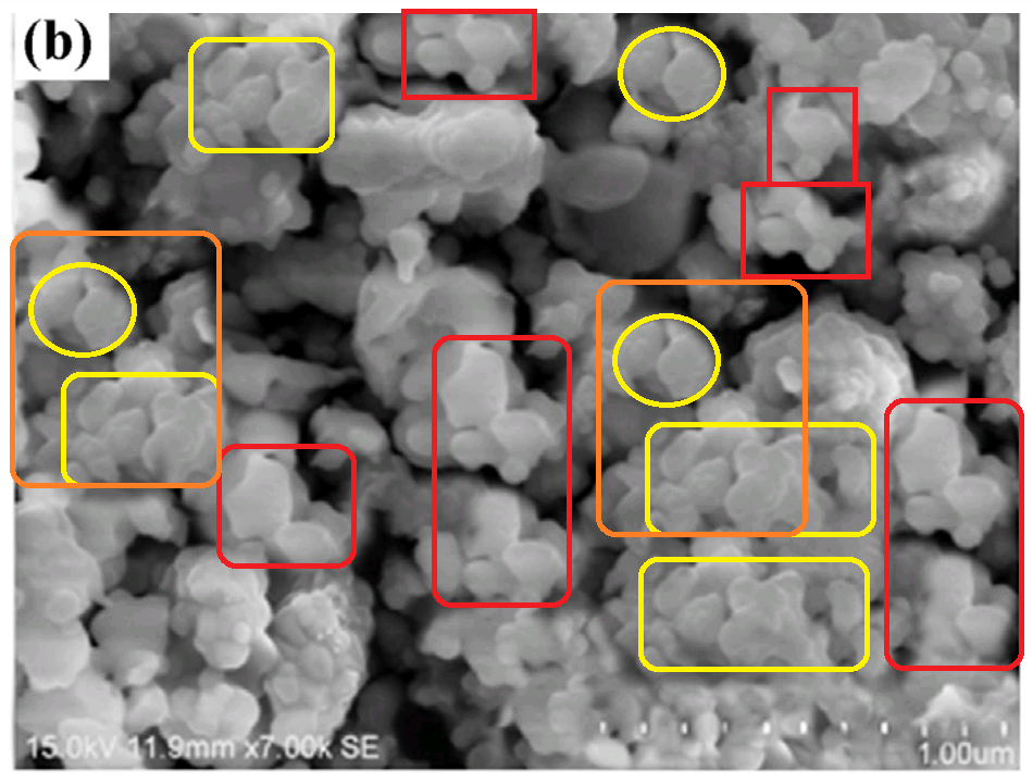
To wet your apatite (as it were), these Scanning Electron Microscope images were presented to the International Conference on Nanoscience and Nanotechnology-2015, before they were published as Fig 3 in Ramya et al. (2015) [19]. Not logged at PubPeer. Here “Mg-HA” is Magnesium-substituted Hydroxy-Apatite.
The failure of other conference delegates to protest such lazy shameless photoshop manipulations by rioting or burning down the venue does not redound to their credit.
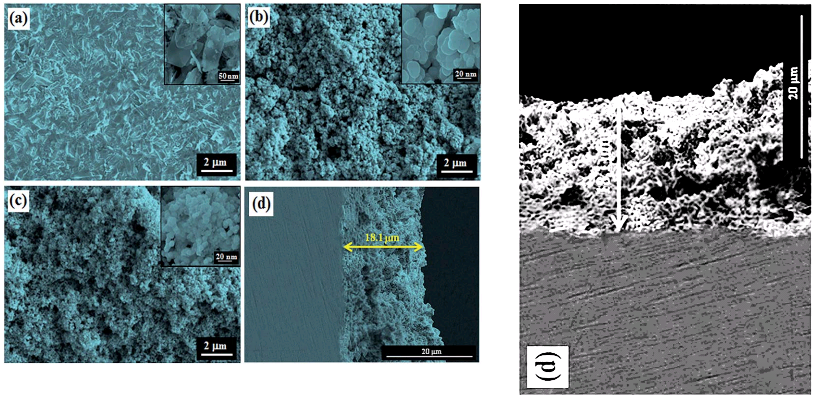
Let us continue with electron microscopy, for this is where Gopi and his colleagues were particularly ill-served by whoever provided them with imagery. Please compare at the left, Fig 3(c) from Gopi et al. (2013b) [3], and at the right, Fig 3(d) from Gopi et al. (2015e) [10].


They depict, respectively, porous Strontium-substituted or Sr/Mg-substituted HA coatings on surgical steel, with polypyrrone or poly(3,4-ethylenedioxythiophene as a kind of primer coat to promote adhesion. The textures are clearly different, while sharing visual motifs in common; one or other did not come directly from the electron microscope.
Figure 7 from Gopi et al. (2015a) [14] was “SEM micrographs of the HA nanoparticles synthesized using malic acid as the chelating agent from the commercial and the naturally available sources; (a) commercial malic acid and (b) apple fruit extract.” I prefer to think that 7(a) was a hommage to Salvador Dali.

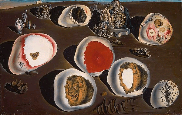
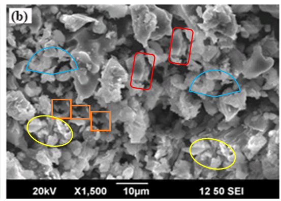
Figure 7(b) is no better.
Figure 7 was reproduced in Gopi et al. (2016b) [22], as Fig 15.14. For Gopi’s work on the chemistry and biology and physical characterisation of modified apatite has led to his recognition as an expert in the perennial genre of “green synthesis of [X] from botanical precursors“. Editors invite him to reprise his discoveries in Review Chapters: in this case, under the title “Chemical and green routes for the synthesis of multifunctional pure and substituted nanohydroxyapatite for biomedical applications”. This is convenient for our purposes as the chapter is a gateway into this section of his oeuvre.
Below, left: Fig 15.17, “SEM micrographs of the HA nanoparticles synthesized using sucrose as a chelating agent from natural and commercially available sources (a) commercial sucrose, (b) pineapple extract, (c) carrot extract, and (d) sugarcane extract.” Who would have expected pineapple- and sugarcane-sourced sucrose to produce such similar nanoparticles, differing only in scale?
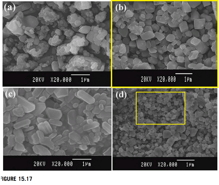
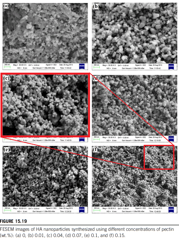
Above, right: Fig 15.19, “FESEM images of HA nanoparticles synthesized using different concentrations of pectin (wt.%): (a) 0, (b) 0.01, (c) 0.04, (d) 0.07, (e) 0.1, and (f) 0.15.” Again, the scale changes but the nanoparticles are otherwise identical.
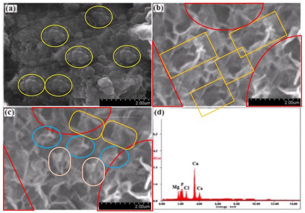
Finally, Fig 15.8 is reproduced from Fig 3 of Gopi et al (2014d) [9]. A sense of unreality pervades these scenes. Is it the ambiguous layering of picture planes, the uncertainty of illusory depth, reminiscent of Matta‘s cosmic dreamscapes? Here are its panels, annotated, not at all phopped:

But some of the tours de force in [9] did not find their way into that Review Paper [22]. Perhaps the authors felt the pressures of space. I feel less constrained in this post, so here are Figs 7(b) and 7(c), “Ca/Sr/Ce–HA nanoparticles synthesized with optimum concentration of Ce3+ (0.1 M) after soaking for … (b) 14 and (c) 21 days in SBF solution”:


Fig 4 consisted of “TEM images of (a) HA, (b) Ca/Sr–HA and (c) Ca/Sr/Ce–HA nanoparticles with optimum concentration (0.1 M) of Ce3+ synthesized by microwave irradiation method”. Below left, 4(b). Below right, 4(a) is compared with Fig 8 from Gopi et al. (2014f), [11], where the particles are derived from banana-skin pectin.


The two images are almost the same, despite their different provenances and scale-bars. Yet they differ along the middle left edge, leading to the melancholy conclusion that one or other has been modified.

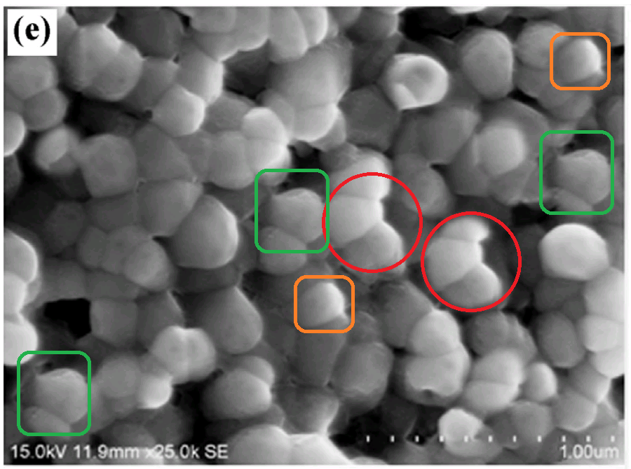
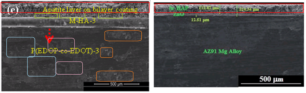
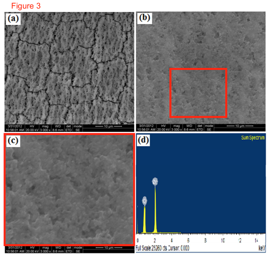
I first encountered Gopi’s group through their work on Nano-Piecrusts: high-resolution Scanning Electron Microscopy cross-sections through a coating of modified hydroxyapatite on a substrate metal (stainless steel, or tungsten, or titanium alloy). The hydroxyapatite is to shield the metal from corrosion or to make it biocompatible as a surgical implant. In Figure 5(i) of Subramani et al. (2016) [24], the pie was “P(EDOP-co-EDOT)/M-HA-3 bilayer coatings on 316L SS”. The internal details of the substrate put Elisabeth Bik in mind of apple crumble pie.
A second pie-slice provided Fig 12(e) in the same paper, “HRSEM cross sectional micrograph of 14 days immersion of P(EDOP-coEDOT)/M-HA-3 bilayer coated 316L SS in SBF solution” (below, left). What commends it to our attention here, apart from the repetitive nature of the polished substrate, is the discovery that the same flaky pastry topping also appeared atop a different substrate, in Fig 3(g) (“Cross-sectional micrograph of Sr-HAP/ZnO duplex-layer-coated AZ91 Mg alloy”) of Gopi et al. (2014b) [6] (below at right).
An intermediate layer of ‘M-HA-3″ gravy also found its way, relabeled as a Polypyrrone-3 primer coat, into Fig 3(d) of [3]. The thickness of the crust looks to me like 3(d) is a shepherd’s pie.

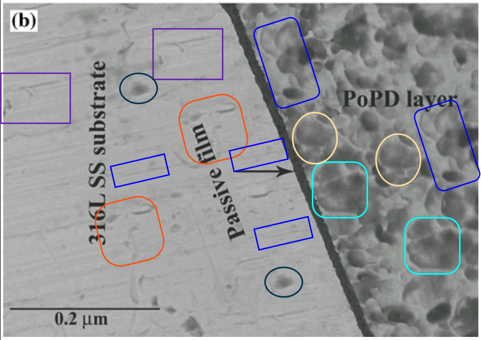
I could easily devote a post to Apatite pie-crust imagery, tracing the tradition back to Fig 11(b) in Gopi et al. (2013) [2], below at left. Perhaps it is an Artist’s Impression of what “SEM cross-sectional micrographs of HAP coating on the post-passivated PoPD-coated 316L SS in Ringer’s solution…. b high magnification for the detection of passive film”would look like, if actually prepared.
Above right is Fig. 2 in Gopi et al. (2015) [13], “Cross-sectional SEM image of 0.5 wt.% H2O2 (optimum concentration) treated AZ91 magnesium alloy after 6 h of immersion in SBF solution”.
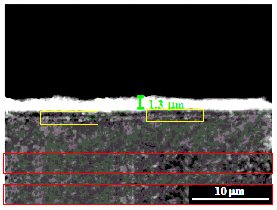
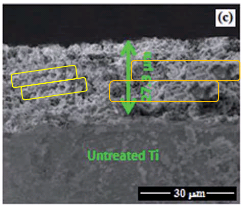
An especially rewarding lode of Piecrust Pastiche is centred on Fig 3(d) of Gopi et al. (2015c) [16], “cross sectional SEM image of CNT/M-HAP coating on Ti”.
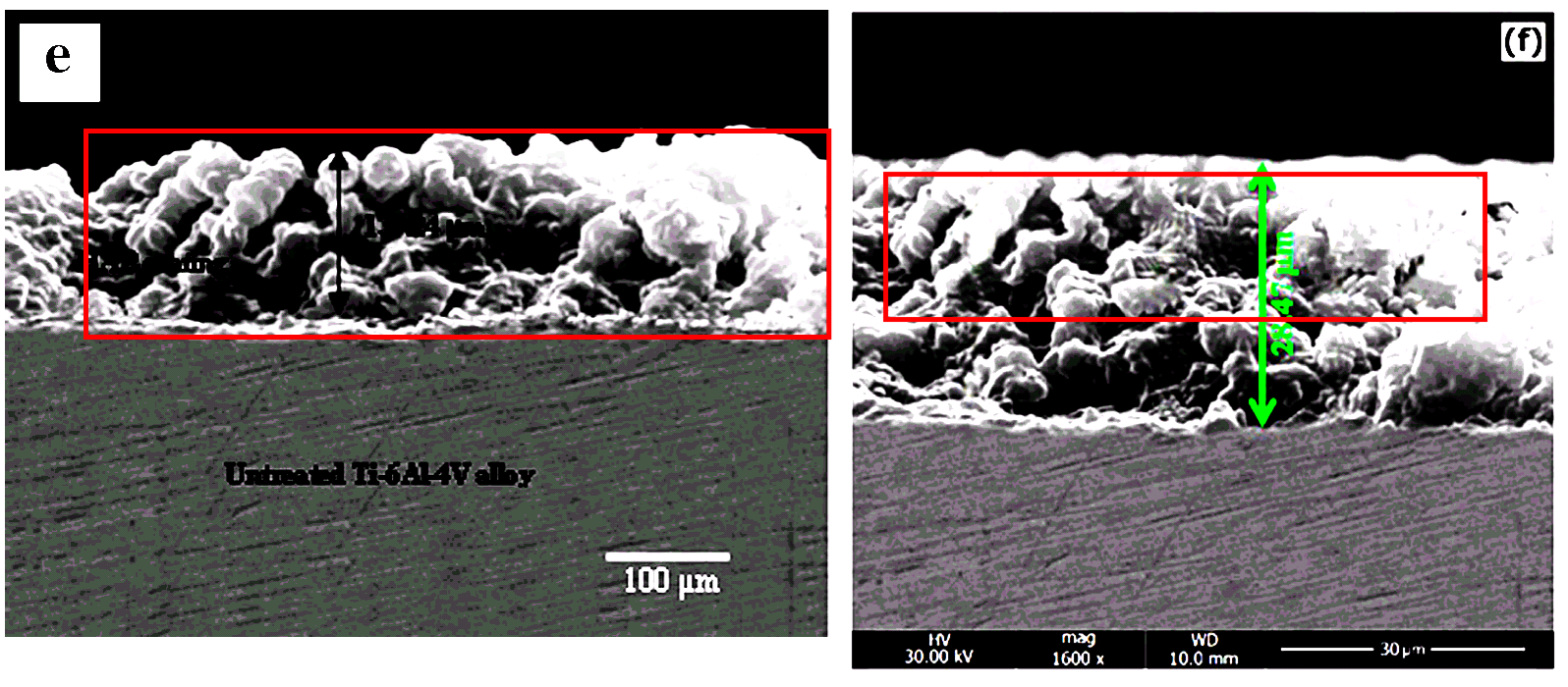
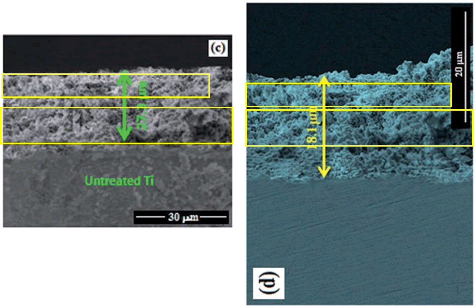
The polished surface of the substrate, with its distinctively recurrent striations, had already appeared in Figures 1(e) of Gopi et al (2014b) [7], and 6(f) of Gopi et al (2014c) [8]. Note that the latter (“the cross sectional image of M-HAP coating on Ti-6Al-4V at 1 s pulse on time and 4 s pulse off time“) has a double-layer crust, one layer being the piecrust from the former (“HAP coating on untreated … Ti–6Al–4V alloy”).
It appears a fourth time in Fig 3(e) of Chozhanathmisra et al. (2016) [20], which is beginning to resemble a Black Forest Gateau, though it has no interpretative legend.
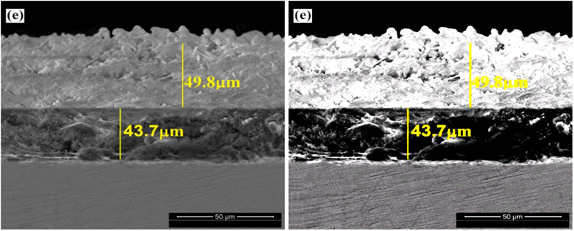
This focus on the curiously striated substrate should not distract us from the topping of 3(d) of [16]. For we find it above a different pie filling – in a flourish of photoshop, with vertical duplications from internal rearrangement – as Fig 4(c) of Gopi et al. (2014) [5] (“cross-sectional image of … the M-HAP coating on untreated Ti”).
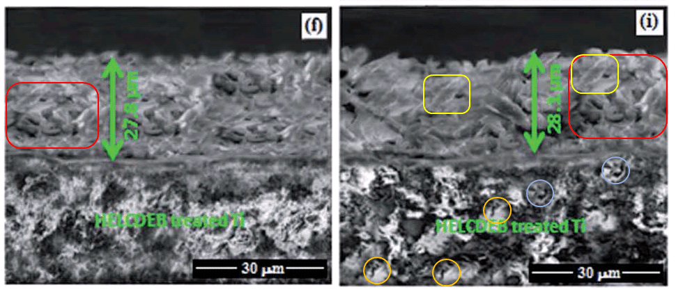
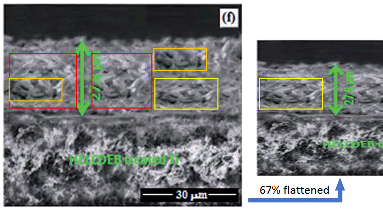
Still with [5], don’t blink, or you will miss the transformation of 4(f) into 4(i) (“cross-sectional image of … the M-HAP coating on 700 keV HELCDEB-treated Ti”). While 4(f) on its own (“cross-sectional image of … the M-HAP coating on 500 keV HELCDEB-treated Ti”) contains fractal graphic motifs deserving of your attention.
Finally, all of Fig 3 from [16] was reprinted as Fig 8.25 in a book chapter, Gopi et al. (2016) [21], edited for Wiley by our old friend Ashutosh Tiwari. None of the Figures reprised in that chapter are credited or accompanied by indications of their non-originality, though this concerns Elsevier copyright lawyers more than it concerns us, and as we will see below, it is the least of the figures’ problems. As if in compensation, the chapter acquired a doubled credit of Gopi’s authorship.
But at this point there is sad news to report… an Erratum has just been published for [24], replacing those iconic piecrusts in Figures 5(i) and 12(e) with less flamboyant photoshoppery. Evidently “they were inadvertently misrepresented” — the outside consultants again. Rather than bicker and argue about who killed who faked them, let us draw a discreet veil over the entire episode, secure in the knowledge that “these corrections do not alter/affect any of the other results or conclusions described in the published manuscript”.
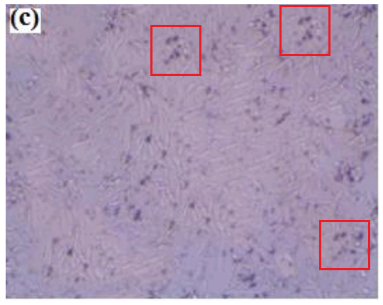
Pastiche pies were not the only casualties of the Erratum, for there is more to biocompatible apatite coatings on metal implants than their cross-sections alone-. To assess their receptiveness to tissue regeneration, Gopi’s team grew fibroblast cell-lines on the surfaces of the materials they synthesized. Then Figure 13 had to be replaced because its panels had already appeared as Fig 9 of [20], identified as “Optical images showing the viability of HOS MG63 cells on Zn-HNT/ M-HA bilayer coating for (a) control (b)1 (c) 4 and (d) 7 days” (rather than as “Optical images showing the viability of HOS MG63 cells on P(EDOP-co-EDOT)/MHA-3 bilayer coatings for (a) 1, (b) 4 and (c) 7 days of incubation”).

They had also been identified as “HOS MG63 cells on S-PEEK/Sr-HAp composite coating for (a) 1, (b) 4 and (c) 7 days…” in Fig 8 of Rajeswari et al. (2014) [12] (below right); and as “HOS MG63 cells on control 4 (a,c,e) … at 1 day, 4 days and 7 days of incubation” in Fig 6 of Gopi et al (2015b) [15] (below right).
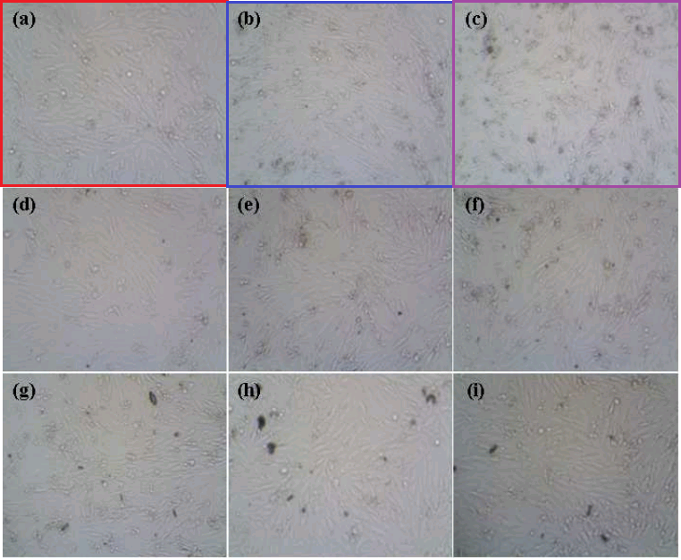

I note in passing that regeneration is particularly good when fibroblast clusters are multiplied with the Photoshop clone stamp. Hence Fig 6 (“optical images of … (c) CNTs/Sr-HAP,… (e) CNTs/Zn-HAP … composite coatings on Ti obtained at 7 days of incubation”) in Gopi et al (2015d) [17]… and then Fig 8.23 in Tiwari’s book [21], for the authors were proud enough of these assisted results to reprint them.

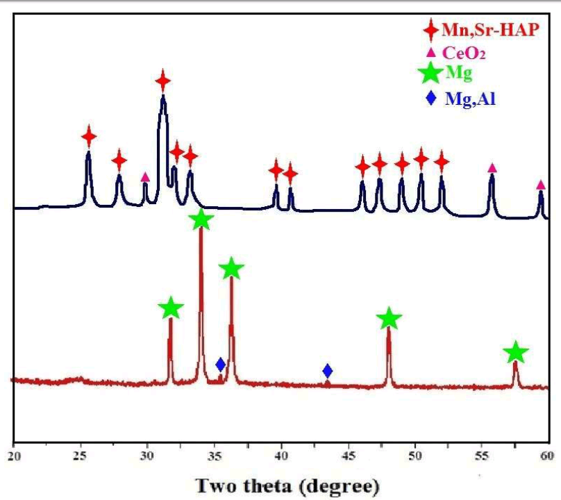
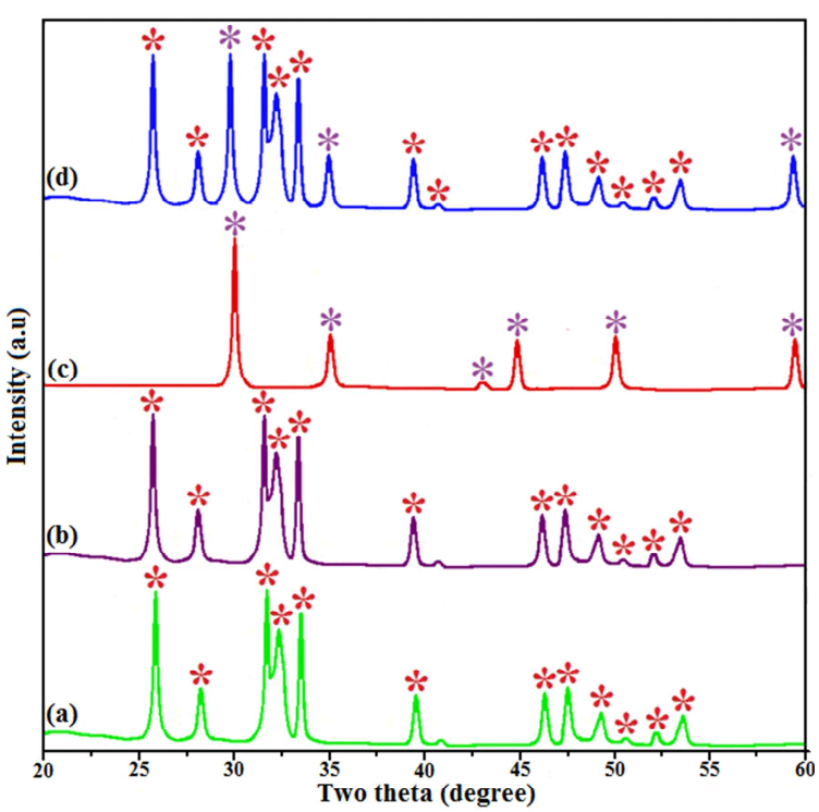
Consolation can be taken from the fact that Fig 3 was not replaced, so presumably the authors have confidence in it. It is an X-Ray Diffraction pattern, artisanal and hand-made in nature, so we know that it was not created by graphical copy-paste. The diffraction peaks burst up from the baseline like Clavaria fungi, like zombies’ fingers thrusting up through the cemetery soil as they dig their way out from their graves.
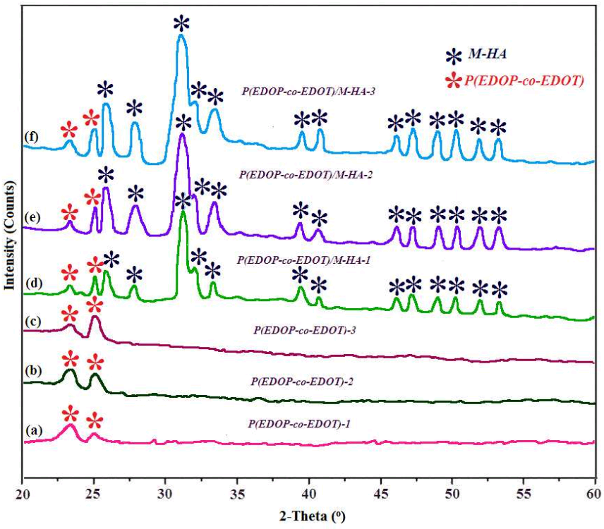
Fig 3 was the most recent in a series of diagrams in which the authors developed a new convention of what XRD patterns should look like, and whether a computer plotter or image file is really a requirement. Earlier zombie-finger exercises include Fig 3 from [12], and Fig 2 from [9].
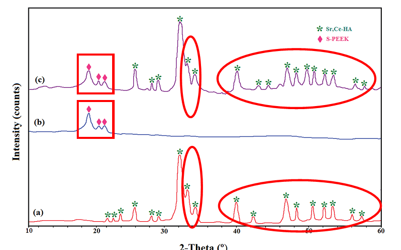
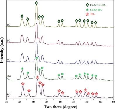
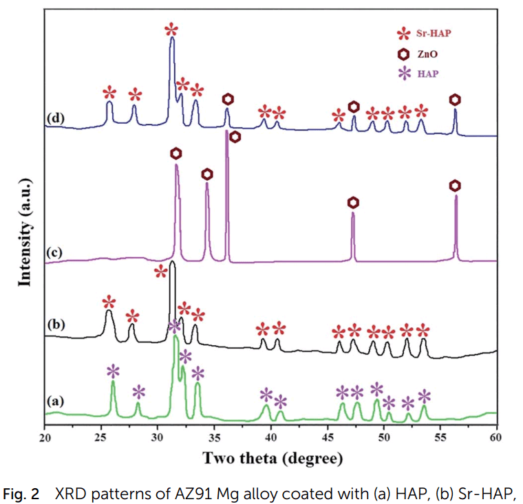
Fig 2 of [6], and Fig 2 of [15]. Was the control profile meant to be a row of palm-trees?
Here by way of contrast are some more traditional diffractograms. At left, an early example: Fig 1 (“XRD patterns of HAP electrodeposited on 316L SS at (a) 1200 mV vs SCE, (b) 1400 mV vs SCE and (c) 1600 mV vs SCE”) from Gopi et al. (2011) [1]. Duplicated stretches are marked. At right, Fig. 2 (“XRD patterns of (a) CNTs/HAP, (b) CNTs/Sr-HAP, (c) CNTs/Mg-HAP (d) CNTs/ZnHAP and (e) CNTs/M-HAP composite coatings”) from [17].
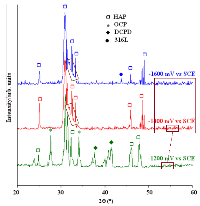
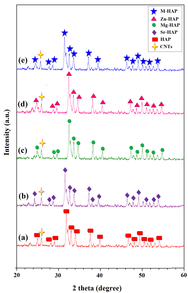
The background of random zigzag noise is the same across the latter patterns, making it easy for one to hide behind another, for little distinguishes them except slight displacements along the Angle axis.

Those experimental outcomes must have been especially satisfying, for they appeared again (as original results) as Fig 8.19 of [21]; and earlier as Fig 2 (“XRD patterns for (a) HAP, (b) M-HAP and (c) CNT/M-HAP coatings”) from [16] – shown below at left.

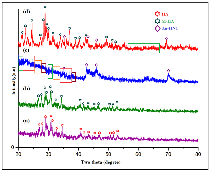

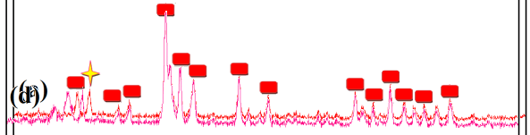
Above right is Fig 2 (“XRD patterns of (a) HA (b) M-HA (c) Zn-HNT and (c) Zn-HNT/ M-HA bilayer coatings”) from [20], where the colors of (a) and (b) are all that distinguish them.
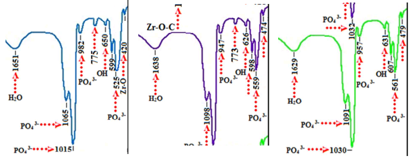
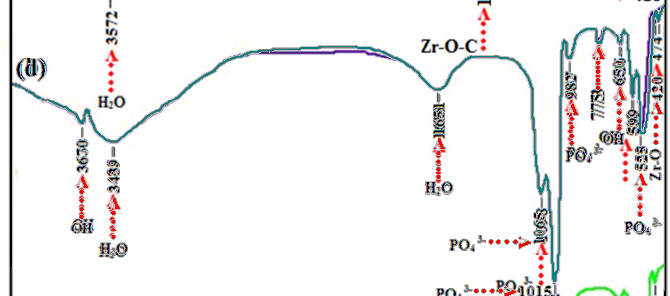
As always, if this post leaves any readers unsatisfied and wanting more, they should review the PubPeer archives. FTIR spectra are another popular tool for characterising freshly-synthesized materials, and for the sake of everyone else, I’ll move on to them.
Fig 1 from Sathishkumar et al. (2016) [23] show “The FT-IR spectra of (a) HAP, (b) Sm/Gd-HAP-I, (c) Sm/Gd-HAP-II and (d) Sm/GdHAP-III coatings”. I am not entirely comfortable with spectra that have overhangs. Were they left out in the sun?
Another source of discomfort are spectra that overlap perfectly over wide bands of frequency. In Fig 1 of [14], it is hard to see light between the low-frequency sections of (a) and (b), apart from the systematic relabelling of every feature.
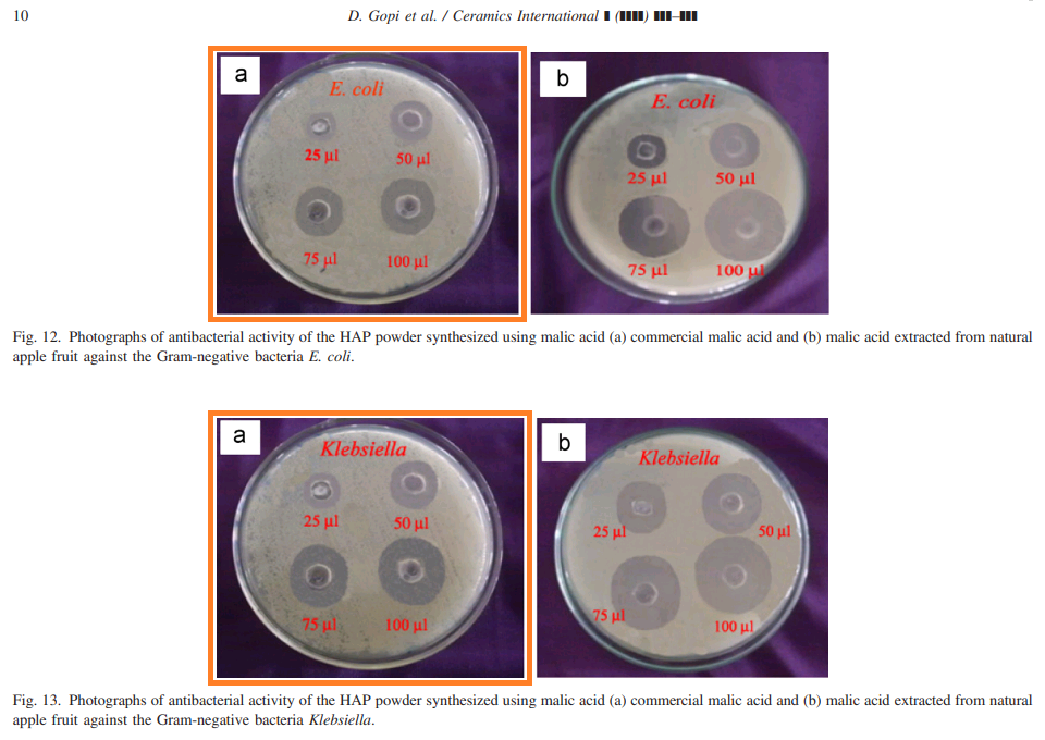
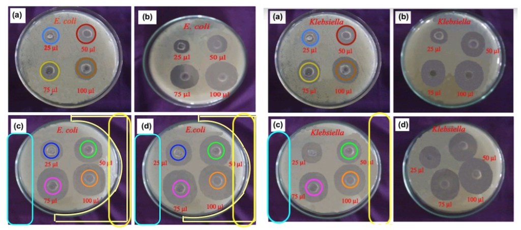
[14] provides a convenient segue to the anti-bacterial properties of these nanoparticles and coatings: a topic not previously addressed, but related to their proposed role as bodily implants. In Figs 12 and 13 (below at left), it is not entirely clear whether the target-practice Petri dishes feature E. coli or Klebsiella. Similar uncertainties plagued the same images when they appeared earlier as Figs 5 and 6 from Gopi et al. (2013c) [4], the nanoparticles derived from grape- or tamarind extract rather than from apples. Below, multicolored annuli adorn the targets to guide the eye and for decoration.
The evidence suggests that the bactericidal performance of these coatings have felt the gentle touch of Appearance-Enhancing Software. The two Petri dishes in Fig 10 of [8] (“Photographs of antimicrobial test results of M-HAP coatings against (a) S. aureus and (b) E. coli (A – M-HAP coating obtained at 4 s pulse on time; and B – M-HAP coating obtained at 4 s pulse off time)”) are more similar than one would expect. This is also true for Fig 6 from [12] (“Antibacterial activity of S-PEEK/Sr-HAp composite coating against (a) E. coli and (b) S. aureus, S-PEEK/Ce-HAp composite coating against (b) E. coli and (c) S. aureus and S-PEEK/Sr,Ce-HAp composite coating against (d) E. coli and (e) S. aureus bacteria”).

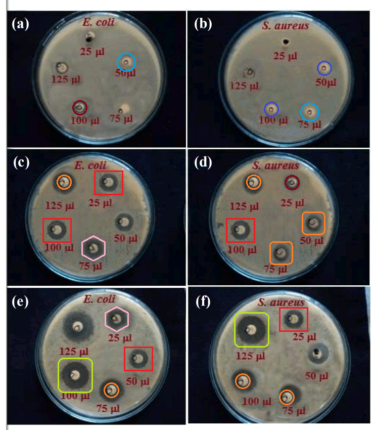
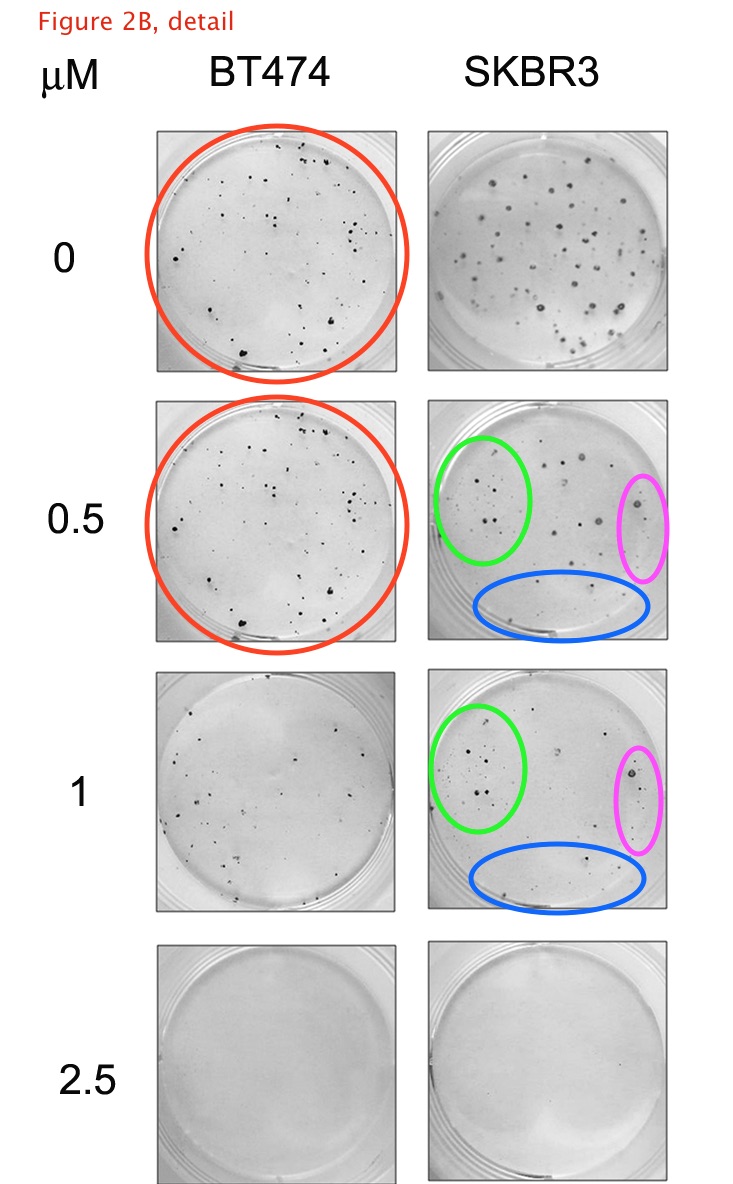
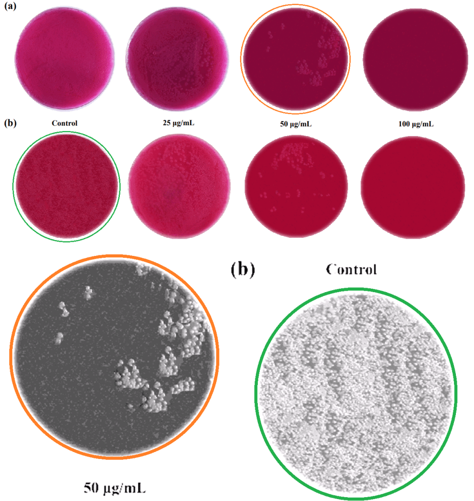
Two last images. Fig 7 of Karthika et al. (2015) [19] showed “Petri dishes with MacConkey agar inoculated with (a) S. aureus and (b) E. coli, showing variable numbers of colonies when supplemented with different amounts of M-HAP/Gel nanocomposite”. My own interpretation is that increasing the quantity of hydroxyapatite / gelatine combination improved the ability of agar to repel the Photoshop clone stamp, as seen more clearly with the magenta tint stripped away and the contrast enhanced.

Acknowledgments
One of the authors D. Gopi acknowledges the major financial support from the Department of Science and Technology, New Delhi, India (DST) and Council of Scientific and Industrial Research, New Delhi, India (CSIR) in the form of major research projects. D. Gopi and L. Kavitha also acknowledge UGC, New Delhi, India for the Research Award (Ref. No. F.30-1/2013(SA-II)/RA-2012-14-NEW-SC-TAM-3240 and F. 30-1/2013(SA-II)/RA-2012-14-NEW-GE-TAM-3228) (2012-2014), respectively.
With all the grants the Periyar team are receiving on the basis of this work, you would think they’d be able to afford more reliable outside consultants.
Sources:
- “A facile electrodeposition of hydroxyapatite onto borate passivated surgical grade stainless steel“, D. Gopi , V. Collins Arun Prakash , L. Kavitha , S. Kannan , P.R. Bhalaji , E. Shinyjoy , J.M.F. Ferreira (2011). Corrosion Science doi: 10.1016/j.corsci.2011.03.018
- “Hydroxyapatite coating on selectively passivated and sensitively polymer-protected surgical grade stainless steel“, D. Gopi , J. Indira , L. Kavitha , J. M. F. Ferreira (2013) Journal of Applied Electrochemistry, doi: 10.1007/s10800-012-0508-z
- “Corrosion protection performance of porous strontium hydroxyapatite coating on polypyrrole coated 316L stainless steel“, D. Gopi, S. Ramya , D. Rajeswari , L. Kavitha (2013b). Colloids and Surfaces B Biointerfaces doi: 10.1016/j.colsurfb.2013.01.065
- “A novel green template assisted synthesis of hydroxyapatite nanorods and their spectral characterization“, D. Gopi, N. Bhuvaneshwari , J. Indira , K. Kanimozhi , L. Kavitha (2013c). Spectrochimica acta. Part A, Molecular and biomolecular spectroscopy doi: 10.1016/j.saa.2013.01.052
- “Investigation on corrosion protection and mechanical performance of minerals substituted hydroxyapatite coating on HELCDEB-treated titanium using pulsed electrodeposition method“, D. Gopi , A. Karthika , D. Rajeswari , L. Kavitha , R. Pramod , Jishnu Dwivedi (2014). RSC Advances doi: 10.1039/c4ra04484c
- “Electrodeposition of a porous strontium-substituted hydroxyapatite/zinc oxide duplex layer on AZ91 magnesium alloy for orthopedic applications“, D. Gopi , N. Murugan , S. Ramya, L. Kavitha (2014b). Journal of Materials Chemistry B doi: 10.1039/c4tb00960f
- “Evaluation of the mechanical and corrosion protection performance of electrodeposited hydroxyapatite on the high energy electron beam treated titanium alloy“, D. Gopi, El-Sayed M. Sherif, D. Rajeswari , L. Kavitha , R. Pramod , Jishnu Dwivedi , S.R. Polak (2014b). Journal of Alloys and Compounds doi: 10.1016/j.jallcom.2014.07.160
- “In vitro biological performance of minerals substituted hydroxyapatite coating by pulsed electrodeposition method“, Dhanaraj Gopi, Arumugam Karthika , Subramani Nithiya , Louis Kavitha (2014c). Materials Chemistry and Physics. doi: 10.1016/j.matchemphys.2013.12.017
- “Strontium, cerium co-substituted hydroxyapatite nanoparticles: Synthesis, characterization, antibacterial activity towards prokaryotic strains and in vitro studies“, D. Gopi, S. Ramya , D. Rajeswari , P. Karthikeyan , L. Kavitha (2014d). Colloids and Surfaces A: Physicochemical and Engineering Aspects doi: 10.1016/j.colsurfa.2014.03.035
- “Development of strontium and magnesium substituted porous hydroxyapatite/poly(3,4-ethylenedioxythiophene) coating on surgical grade stainless steel and its bioactivity on osteoblast cells“, D. Gopi, S. Ramya , D. Rajeswari , M. Surendiran , L. Kavitha (2014e). Colloids and Surfaces B Biointerfaces doi: 10.1016/j.colsurfb.2013.10.011.
- “Novel banana peel pectin mediated green route for the synthesis of hydroxyapatite nanoparticles and their spectral characterization“, D. Gopi, K. Kanimozhi , N. Bhuvaneshwari , J. Indira , L. Kavitha (2014f). Spectrochimica acta. Part A, Molecular and biomolecular spectroscopy doi: 10.1016/j.saa.2013.09.034
- “Investigation of anticorrosive, antibacterial and in vitro biological properties of a sulphonated poly(etheretherketone)/strontium, cerium co-substituted hydroxyapatite composite coating developed on surface treated surgical grade stainless steel for orthopedic applications“, D. Rajeswari , D. Gopi, S. Ramya , L. Kavitha (2014). RSC Advances doi: 10.1039/c4ra12207k
- “Evaluation of biodegradability of surface treated AZ91 magnesium alloy in SBF solution“, D. Gopi , P.R. Bhalaji , S. Ramya , L. Kavitha (2015). Journal of Industrial and Engineering Chemistry doi: 10.1016/j.jiec.2014.08.019
- “Novel malic acid mediated green route for the synthesis of hydroxyapatite particles and their spectral characterization“, D. Gopi, N. Bhuvaneshwari , L. Kavitha , S. Ramya (2015a). Ceramics International doi: 10.1016/j.ceramint.2014.10.156
- “Ball flower like manganese, strontium substituted hydroxyapatite/cerium oxide dual coatings on the AZ91 Mg alloy with improved bioactive and corrosion resistance properties for implant applications“, D. Gopi , N. Murugan , S. Ramya , E. Shinyjoy , L. Kavitha (2015b). RSC Advances doi: 10.1039/c5ra03432a
- “Single walled carbon nanotubes reinforced mineralized hydroxyapatite composite coatings on titanium for improved biocompatible implant applications“, D. Gopi , E. Shinyjoy , A. Karthika , S. Nithiya , L. Kavitha , D. Rajeswari , Tingting Tang (2015c). RSC Advances doi: 10.1039/c5ra04382d
- “Influence of ionic substitution in improving the biological property of carbon nanotubes reinforced hydroxyapatite composite coating on titanium for orthopedic applications“, D. Gopi , E. Shinyjoy , L. Kavitha (2015d). Ceramics International doi: 10.1016/j.ceramint.2014.12.114
- “Fabrication of divalent ion substituted hydroxyapatite/gelatin nanocomposite coating on electron beam treated titanium: mechanical, anticorrosive, antibacterial and bioactive evaluations“, A. Karthika , L. Kavitha , M. Surendiran , S. Kannan , D. Gopi (2015). RSC Advances doi: 10.1039/c5ra05624a
- “Corrosion and biodegradability evaluation of magnesium substituted porous hydroxyapatite/polyethylene dioxythiophene bilayer coating on 316l stainless steel for orthopaedic applications”, S. Ramya, D. Rajeswari, M. Palanisamy, D. Gopi , L. Kavitha (2015). International Journal of ChemTech Research.
- “Development of zinc-halloysite nanotube/minerals substituted hydroxyapatite bilayer coatings on titanium alloy for orthopedic applications“, M. Chozhanathmisra , S. Ramya , L. Kavitha , D. Gopi (2016). Colloids and Surfaces A: Physicochemical and Engineering Aspects doi: 10.1016/j.colsurfa.2016.10.018
- “Carbon Nanotubes-reinforced Bioceramic Composite: An Advanced Coating Material for Orthopedic Applications“, D. Gopi , D. Gopi , E. Shinyjoy , L. Kavitha , D. Rajeswari (2016). Advanced Composite Materials doi: 10.1002/9781119242666.ch8
- “Chemical and green routes for the synthesis of multifunctional pure and substituted nanohydroxyapatite for biomedical applications“, Dhanaraj Gopi , Louis Kavitha , Subramanian Ramya , Durairajan Rajeswari (2016b). Engineering of Nanobiomaterials doi: 10.1016/b978-0-323-41532-3.00015-4
- “Tailoring the Sm/Gd-Substituted Hydroxyapatite Coating on Biomedical AISI 316L SS: Exploration of Corrosion Resistance, Protein Profiling, Osteocompatibility, and Osteogenic Differentiation for Orthopedic Implant Applications“, Saravanan Sathishkumar , Kavitha Louis , Elangomannan Shinyjoy , Dhanaraj Gopi (2016). Industrial & Engineering Chemistry Research doi: 10.1021/acs.iecr.5b04329
- “Fabrication of Minerals Substituted Porous Hydroxyapaptite/Poly(3,4-ethylenedioxy pyrrole-co-3,4-ethylenedioxythiophene) Bilayer Coatings on Surgical Grade Stainless Steel and Its Antibacterial and Biological Activities for Orthopedic Applications“, Ramya Subramani , Shinyjoy Elangomannan , Kavitha Louis , Soundarapandian Kannan, Dhanaraj Gopi (2016). ACS Applied Materials & Interfaces doi: 10.1021/acsami.6b01795.

Donate!
If you are interested to support my work, you can leave here a small tip of $5. Or several of small tips, just increase the amount as you like (2x=€10; 5x=€25). Your generous patronage of my journalism, however small it appears to you, will greatly help me with my legal costs.
5.00 €


Shorter G Dopey (mis-spellings, my own:
“We have no expertise in biology.”
LikeLike
It is really disturbing that most of journals (not only ACS) prefer to publish corrections instead of retracting papers with clearly manipulated data. Even if small piece of data was manipulated by authors the rest of their paper can not be trusted. These papers must be retracted.
. May be they are afraid of Retraction Watch publications? In this case one need to create “No Retraction Watch” for balance. It should be ok for journals to retract paper but completely wrong not to retract. Now it is all upside down.
LikeLike
It stands to reason that All Crap Science clings to disclaimers like: “These corrections do not affect any of the major conclusions of the paper.” They may actually be right. If the major conclusion is that the paper is crap, and so are the “researchers” that fabricated it, then a definitive demonstration of their collective crappiness merely adds a mite of confidence to the obvious. Nevertheless, kudos to SC for outing these wastrels. Shining a strong light into their cockroach corner will at least force them to scurry off for a few minutes. They should credit those felt-tip spectra to “Shaky Shiva”.
LikeLike
Bad day for science, just makes me sad.
LikeLike
Dr Gopi spreads his expertise widely. Here delving into Fabrication of a pH responsive DOX conjugated PEGylated palladium nanoparticle mediated drug delivery system: an in vitro and in vivo evaluation:
LikeLike
Co-author Raju Vivek came to Pubpeer (or was sent) to defend Fig 8 in HER2 Targeted Breast Cancer Therapy with Switchable “Off/On” Multifunctional “Smart” Magnetic Polymer Core–Shell Nanocomposites.
“Secondly, coming to the Figure 8, histology experiment, we used the different tissues of the control as well as treated animals to prove the nontoxicity of composite which we selected. Furthermore, the cellular architecture of organs like kidney and Liver or any other organs used in the study may be similar. The staining intensity is differ from one another. Therefore, there is possibilities of look like similarity cells of the organ images but its totally different kinds of the organs used to done histology.”
People were not convinced.
LikeLike
Smart rose flower like bioceramic/metal oxide dual layer coating with enhanced anti-bacterial, anti-cancer, anti-corrosive and biocompatible properties for improved orthopedic applications is another one that ended up on the cutting-room floor. It had photocopied FTIR spectra…
LikeLike
…AND fake zombie-finger XRDs…


LikeLike
…AND another selection of SEMs where the effect of longer deposition time was to enlarge the scale of the deposit.
Well done RSC Advances.
LikeLike
One more out-take! No room in the main text for Carbon Nanofiber/Polycaprolactone/Mineralized Hydroxyapatite Nanofibrous Scaffolds for Potential Orthopedic Applications
. Includes zombie fingers…
AND downward-zombie-finger FTIRs.
LikeLike
Pingback: Deliferate misteaks by Tradional Chinese Medicine – For Better Science
Pingback: Linköping haunted by fake spectra – For Better Science
One more best research from his best work.
https://timesofindia.indiatimes.com/city/coimbatore/VC-seeks-explanation-from-prof-accused-of-sexual-assault/articleshow/51149303.cms
LikeLike
From 2016:
“The vice-chancellor of Periyar University on Thursday issued a memo to professor D Gopi, accused of sexual and physical harassment of a few Ph.D students, seeking an explanation from him within the next 10 days. The move comes after the committees, which looked into the issue, in their report said the professor was prima facie guilty in the case.
“Based on the reports, I have issued the memo, seeking an explanation from professor Gopi,” said vice-chancellor C Swaminathan. “Action will be initiated against him after receiving the explanation letter,” he further said.
On February 8, a group of seven Ph.D students, including three girls, had petitioned the vice-chancellor against the chemistry professor, Gopi, saying he had abused and harassed them sexually as well as physically.
In their petition, the students said the professor made male students to do household chores such as washing clothes, room cleaning and paying mobile phone bills, while he sexually harassed girl students. The professor also allegedly made the girl students to take care of his new born twins when he went out with his wife for shopping, besides forcing them to attend to his wife at hospital when she was admitted for delivery.
Upon receiving the petition, Swaminathan constituted a four-member committee comprising the dean of school of physical sciences, V Krishnakumar, professors V Sangeetha (English department) and K Dhanalakshmi (Education) and an external professor from Bharathiyar University, Coimbatore, K Dharmaraj (Chemistry department).
The girls also lodged a compliant with anti-sexual harassment cell at the Periyar University against the professor. The cell also conducted an inquiry in this regard.
The two committees had on Tuesday submitted their reports to the vice-chancellor, stating that they prima facie found the professor guilty.”
LikeLike
He got ‘Tamil Nadu Scientist Award’ for his great research work
https://www.thehindu.com/news/cities/Coimbatore/periyar-university-professor-gets-tamil-nadu-scientist-award/article19822868.ece
LikeLike
Oh yes, sexual harassment must be rewarded.
“D. Gopi, Professor of Chemistry, Periyar University, has been honoured with ‘Tamil Nadu Scientist Award’ for 2013 in recognition of his contributions in the field of medical sciences.
The award has been instituted by the Tamil Nadu State Council for Science and Technology (TNSCST).
Mr. Gopi received the award from K. P. Anbalagan, State Higher Education Minister, in the presence of Sunil Paliwal, Higher Education Secretary, and R. Srinivasan, Member Secretary of Tamil Nadu State Council for Science and Technology, at a function held in Chennai recently
LikeLike
What achievement ! Professor Gopi got Total Grant Mobilized : 2.25 Crore from Government of India for his fake (data fabrication factory) research.. OMG
Project Details
Click to access dr.d.gopi-bio-data.pdf
LikeLike
Pingback: Thomas Webster to save the world with COVID-19 nanoparticles – For Better Science
Pingback: Whitewashed by ACS and Elsevier, Zboril sues Olomouc Dean for “bullying” – For Better Science
Pingback: Bad Choices in Dresden – For Better Science
Pingback: The One-Man Papermill – For Better Science
Pingback: Longwei Yin corrections, or what’s the point anyway – For Better Science